Deposition Date
2024-05-03
Release Date
2025-01-01
Last Version Date
2025-04-23
Entry Detail
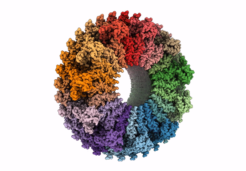
PDB ID:
8ZDS
Keywords:
Title:
Structure of the Salmonella flagellar MS-ring with C11 symmetry applied
Biological Source:
Source Organism(s):
Expression System(s):
Method Details:
Experimental Method:
Resolution:
3.10 Å
Aggregation State:
PARTICLE
Reconstruction Method:
SINGLE PARTICLE