Deposition Date
2024-04-18
Release Date
2025-04-23
Last Version Date
2025-09-24
Entry Detail
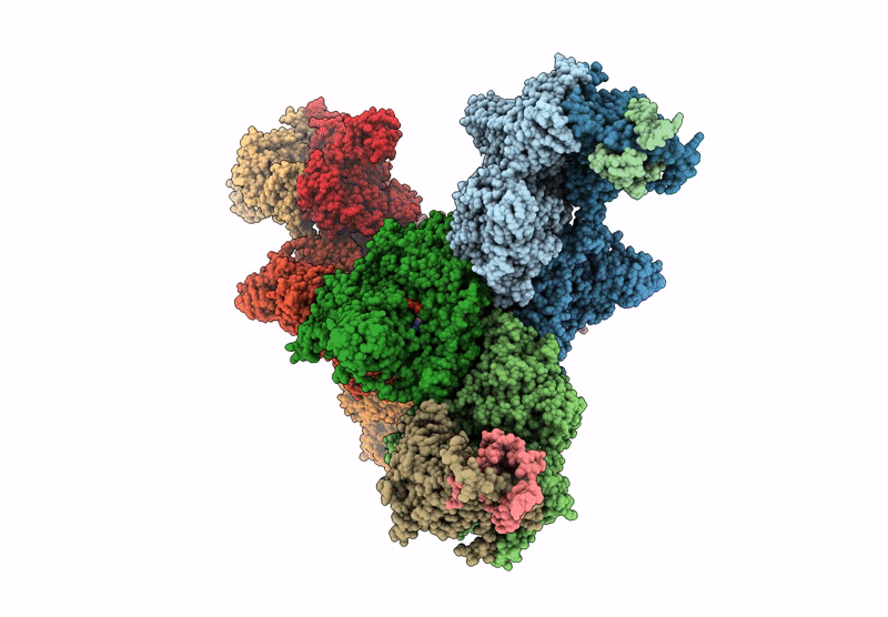
PDB ID:
8Z5T
Keywords:
Title:
human phosphorylase kinase - phosphorylation and Ca2+ bound state
Biological Source:
Source Organism:
Homo sapiens (Taxon ID: 9606)
Host Organism:
Method Details:
Experimental Method:
Resolution:
3.74 Å
Aggregation State:
PARTICLE
Reconstruction Method:
SINGLE PARTICLE