8Z0G image
Deposition Date 2024-04-09
Release Date 2024-12-25
Last Version Date 2024-12-25
Entry Detail
PDB ID:
8Z0G
Title:
Crystal structure of NeIle complexed with isoleucine
Biological Source:
Source Organism:
Host Organism:
Method Details:
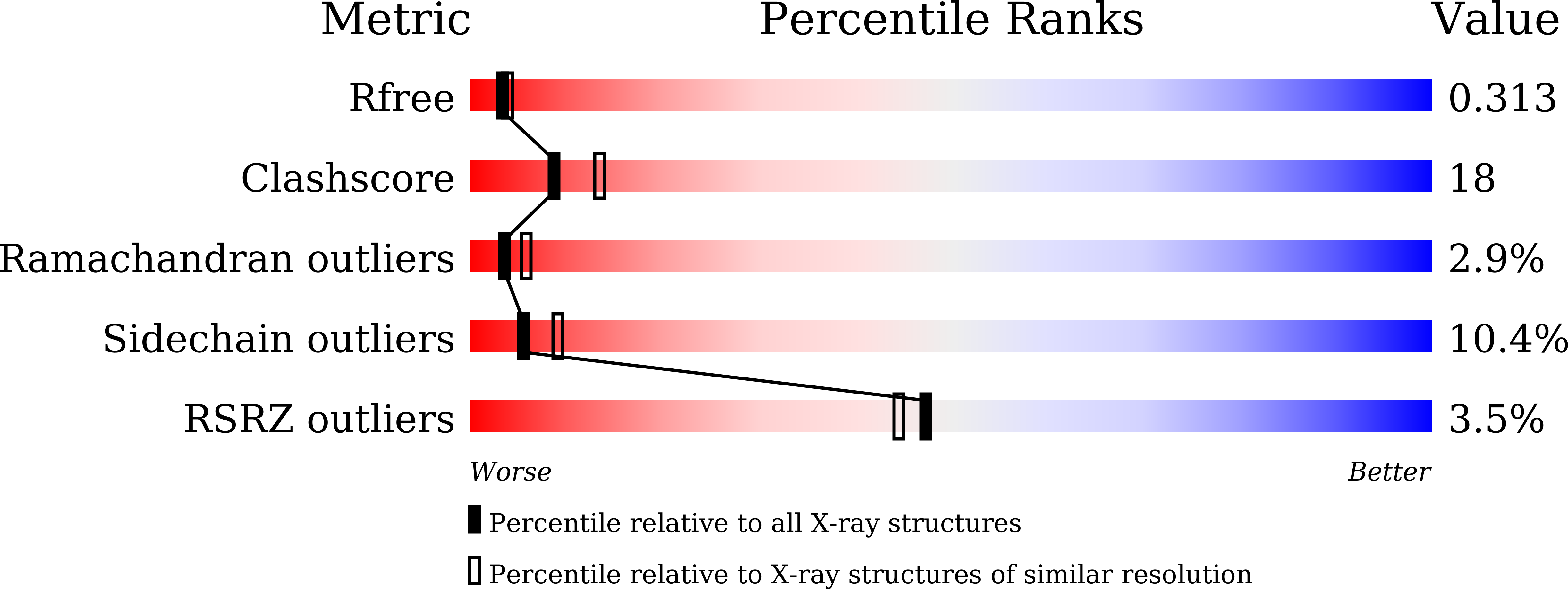
Experimental Method:
Resolution:
2.65 Å
R-Value Free:
0.30
R-Value Work:
0.23
R-Value Observed:
0.23
Space Group:
P 1 21 1
Macromolecular Entities
Structures with similar UniProt ID
Protein Blast
Polymer Type:polypeptide(L)
Molecule:Leu/Ile/Val-binding protein,Leu/Ile/Val-binding protein,mNeonGreen
Chain IDs:A
Chain Length:595
Number of Molecules:1
Biological Source:Escherichia coli O157:H7, Branchiostoma lanceolatum
Primary Citation
NeIle, a Genetically Encoded Indicator for Branched-Chain Amino Acids Based on mNeonGreen Fluorescent Protein and LIVBP Protein.
ACS Sens 9 5135 5147 (2024)
PMID: 39400357 DOI: 10.1021/acssensors.4c01055

Abstact

Branched-chain amino acids (BCAAs) play an important role in the functioning of mammalian cells and the central nervous system. However, available genetically encoded indicators for BCAAs are based on Förster resonance energy transfer and have a limited dynamic range. We developed a single fluorescent protein-based sensor for BCAAs, called NeIle, which is composed of circularly permutated mNeonGreen protein inserted into the leucine-isoleucine-valine binding protein (LIVBP) from Escherichia coli bacteria. In solution, the NeIle indicator displayed a positive fluorescence response to adding isoleucine, leucine, and valin amino acids with high ΔF/F dynamic ranges of 27-, 19-, and 11-fold and the corresponding affinity values of 5.0, 2.9, and 75 mM, respectively. The spectral and biochemical properties of the NeIle indicator were characterized in solution. We characterized the brightness of the NeIle indicator in living mammalian cells, including cultured neurons. Using the NeIle indicator, we successfully visualized the dynamics of isoleucine transients in different organelles of mammalian cells. We obtained and analyzed the X-ray crystal structure of the NeIle indicator in an isoleucine-bound state. Structure-guided directed mutagenesis of the NeIle indicator revealed the basis of its fluorescence response and selectivity to isoleucine.

Legend

Protein

Chemical

Disease

Primary Citation of related structures