8YXB image
Entry Detail
PDB ID:
8YXB
Title:
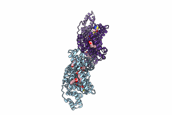
Crystal structure of the HSA complex with ceftriaxone and myristate
Biological Source:
Source Organism:
Host Organism:
PDB Version:
Deposition Date:
2024-04-02
Release Date:
2024-08-07
Method Details:
Experimental Method:
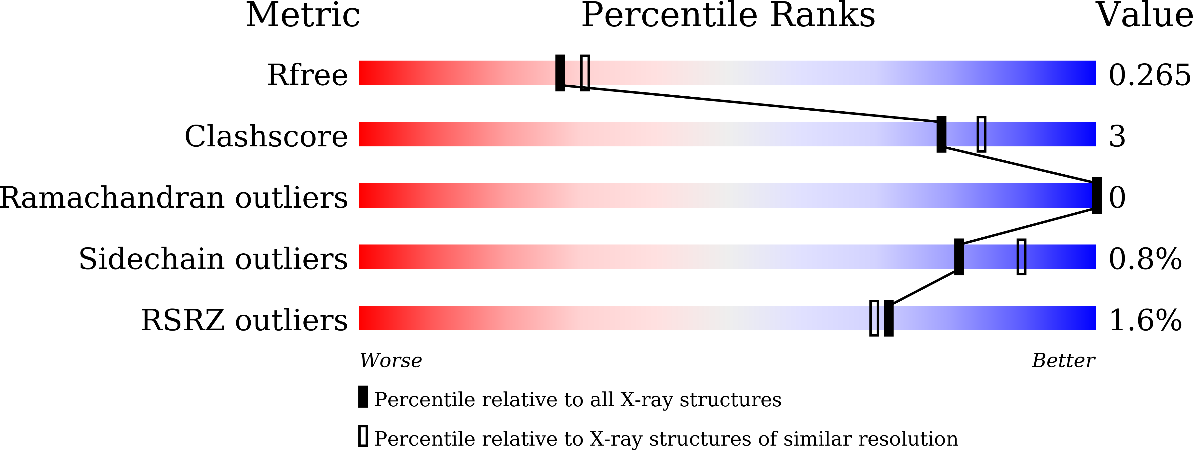
Resolution:
2.20 Å
R-Value Free:
0.26
R-Value Work:
0.23
R-Value Observed:
0.23
Space Group:
P 1
Macromolecular Entities
Polymer Type:polypeptide(L)
Description:Serum albumin
Chain IDs:A, B
Chain Length:585
Number of Molecules:2
Biological Source:Homo sapiens
Primary Citation
Interaction of Cephalosporins with Human Serum Albumin: A Structural Study.
J.Med.Chem. 67 14175 14183 (2024)
PMID: 39083648 DOI: 10.1021/acs.jmedchem.4c00983

Abstact

Modification of the R1 and R2 side chain structures has been used as the main strategy to expand the spectrum of cephalosporins and impart resistance to hydrolysis by β-lactamases. These structural modifications also result in a wide range of plasma protein binding, especially with human serum albumin (HSA). Here, we determined the crystal structures of the HSA complexes with two clinically important cephalosporins, ceftriaxone and cefazolin, and evaluated the binding of cephalosporin to HSA by susceptibility testing and competitive binding assay. Ceftriaxone and cefazolin bind to subdomain IB of HSA, and their cephem core structures are recognized by Arg117 of HSA. Tyr161 of HSA changes its rotamer depending on the cephalosporin, resulting in alterations of the cavity shape occupied by the R2 side chain of cephalosporins. These findings provide structural insight into the mechanisms underlying the HSA binding of cephalosporins.

Legend

Protein

Chemical

Disease

Primary Citation of related structures