
Deposition Date
2024-03-29
Release Date
2025-03-19
Last Version Date
2025-03-19
Entry Detail
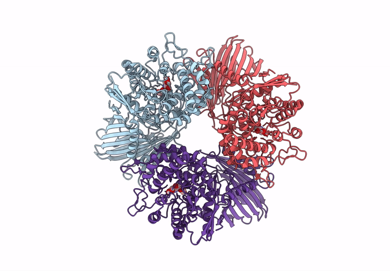
PDB ID:
8YVR
Keywords:
Title:
Crystal structure of GH65 alpha-1,2-glucosidase from Flavobacterium johnsoniae in complex with 1-deoxynojirimycin
Biological Source:
Source Organism(s):
Flavobacterium johnsoniae UW101 (Taxon ID: 376686)
Expression System(s):
Method Details:
Experimental Method:
Resolution:
1.90 Å
R-Value Free:
0.27
R-Value Work:
0.25
Space Group:
C 1 2 1
