8YVB image
Entry Detail
PDB ID:
8YVB
Title:
Crystal structure of Caenorhabditis elegans ZIM-1 ZF1-2-CTD domain in complex with Chromosome II/III pairing center
Biological Source:
Source Organism:
Host Organism:
PDB Version:
Deposition Date:
2024-03-28
Release Date:
2024-12-11
Method Details:
Experimental Method:
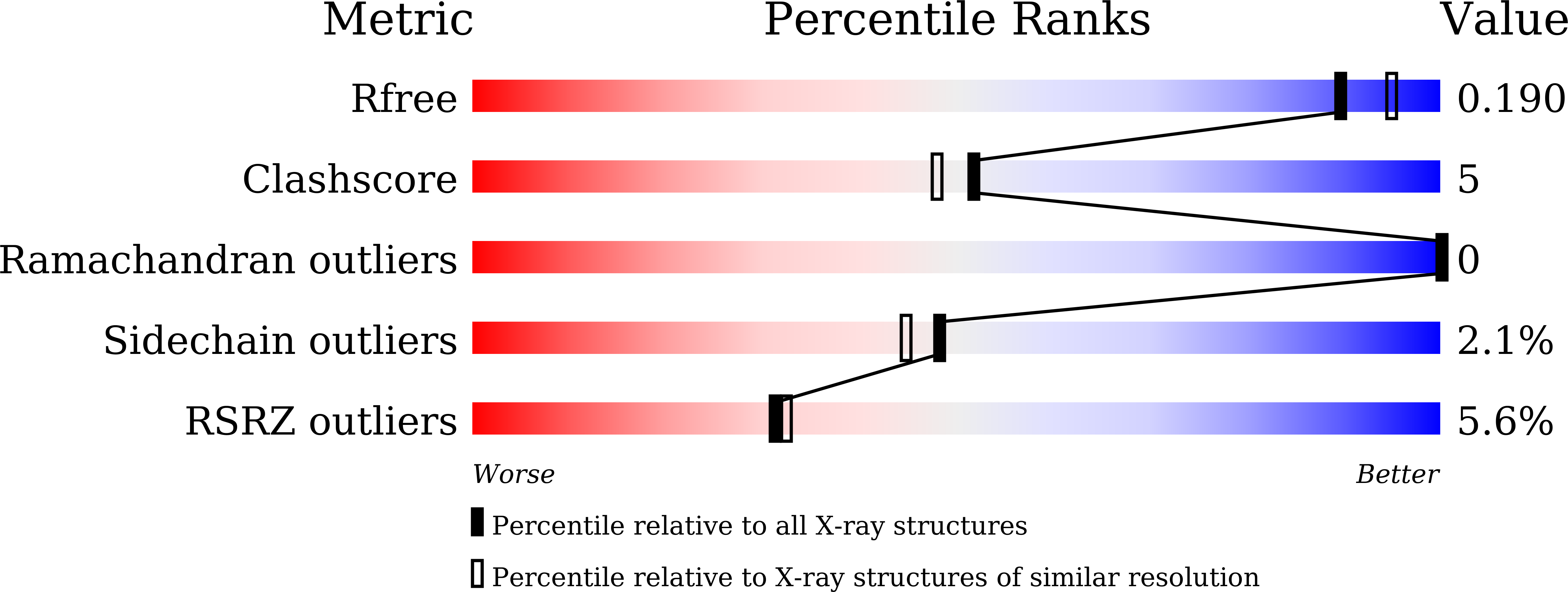
Resolution:
1.90 Å
R-Value Free:
0.23
R-Value Work:
0.18
R-Value Observed:
0.19
Space Group:
C 2 2 21
Macromolecular Entities
Polymer Type:polypeptide(L)
Description:C2H2-type domain-containing protein
Chain IDs:A
Chain Length:128
Number of Molecules:1
Biological Source:Caenorhabditis elegans
Polymer Type:polydeoxyribonucleotide
Description:DNA (5'-D(*TP*TP*GP*GP*TP*CP*TP*GP*CP*TP*A)-3')
Chain IDs:B
Chain Length:11
Number of Molecules:1
Biological Source:Caenorhabditis elegans
Polymer Type:polydeoxyribonucleotide
Description:DNA (5'-D(P*AP*GP*CP*AP*GP*AP*CP*CP*AP*A)-3')
Chain IDs:C
Chain Length:10
Number of Molecules:1
Biological Source:Caenorhabditis elegans
Ligand Molecules
Primary Citation
Structural basis for C. elegans pairing center DNA binding specificity by the ZIM/HIM-8 family proteins.
Nat Commun 15 10355 10355 (2024)
PMID: 39609407 DOI: 10.1038/s41467-024-54548-9

Abstact

Pairing center (PC) on each chromosome of Caenorhabditis elegans is crucial for homolog pairing and initiating synapsis. Within each PC, clusters of 11/12 bp DNA motif recruit one of four paralogous meiosis-specific proteins: ZIM-1, ZIM-2, ZIM-3, or HIM-8. However, the mechanistic basis underlying the specificity of ZIM/HIM-8-PC DNA interaction remains elusive. Here, we describe crystal structures of HIM-8, ZIM-1 and ZIM-2 DNA binding domains (ZF1, ZF2 and CTD) in complex with their cognate PC DNA motifs, respectively. These structures demonstrated the ZF1-2-CTD folds as an integrated structural unit crucial for its DNA binding specificity. Base-specific DNA-contacting residues are exclusively distributed on ZF1-2 and highly conserved. Furthermore, the CTD potentially contributes to the conformational diversity of ZF1-2, imparting binding specificity to distinct PC DNA motifs. These findings shed light on the mechanism governing PC DNA motif recognition by ZIM/HIM-8 proteins, suggesting a co-evolution relationship between PC DNA motifs and ZF1-2-CTD in shaping the specific recognition.

Legend

Protein

Chemical

Disease

Primary Citation of related structures