8YTS image
Deposition Date 2024-03-26
Release Date 2024-04-24
Last Version Date 2025-07-30
Entry Detail
PDB ID:
8YTS
Title:
The structure of the cytochrome c546/556 from Thioalkalivibrio paradoxus with unusual UV-Vis spectral features at atomic resolution
Biological Source:
Source Organism:
Method Details:
Experimental Method:
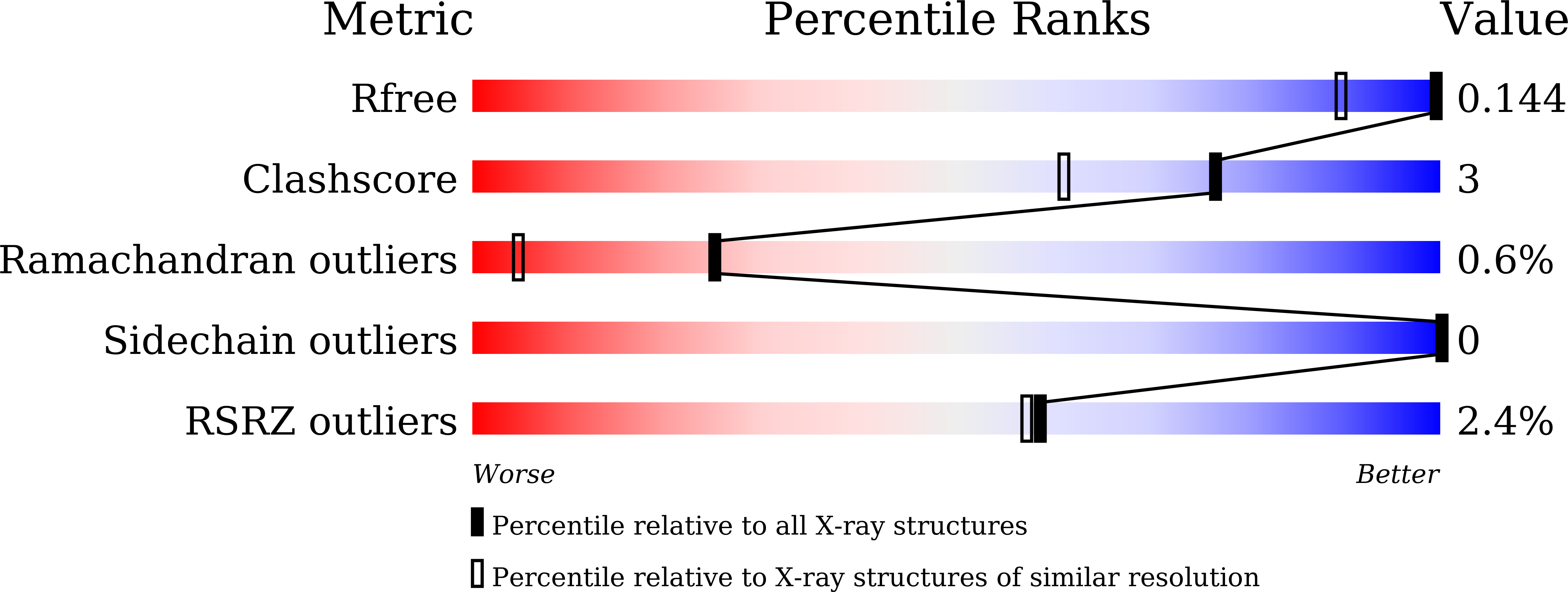
Resolution:
1.15 Å
R-Value Free:
0.14
R-Value Work:
0.11
R-Value Observed:
0.11
Space Group:
P 21 21 21
Macromolecular Entities
Structures with similar UniProt ID
Protein Blast
Polymer Type:polypeptide(L)
Molecule:Cytochrome C
Gene (Uniprot):THITH_15890
Chain IDs:A, B
Chain Length:105
Number of Molecules:2
Biological Source:Thioalkalivibrio paradoxus ARh 1
Ligand Molecules
Primary Citation
Relationship between the structure and physicochemical properties of cytochrome c 546/556 from the bacterium Thioalkalivibrio paradoxus ARh1.
Biochem.Biophys.Res.Commun. 778 152340 152340 (2025)
PMID: 40669334 DOI: 10.1016/j.bbrc.2025.152340

Abstact

An interplay between the structural and physicochemical properties of the monoheme cytochromes of type c has been extensively studied. However new proteins belonging to this diverse family continue to reveal some novel and unique features. Here, we present the 1.15 Å structure of the low-potential cytochrome c546/556 from the bacterium Thioalkalivibrio paradoxus ARh1, which exhibits the prominent splitting of the Q bands in UV-visible spectra even at room temperature. The data obtained suggest that two conformations of the propionate 7 of the heme are responsible for the splitting of the Q bands. We propose that the degree of the splitting of the Q bands is correlated with the conformational lability of the heme propionates.

Legend

Protein

Chemical

Disease

Primary Citation of related structures
Feedback Form
Name
Email
Institute
Feedback