Deposition Date
2024-03-25
Release Date
2024-12-11
Last Version Date
2025-02-12
Entry Detail
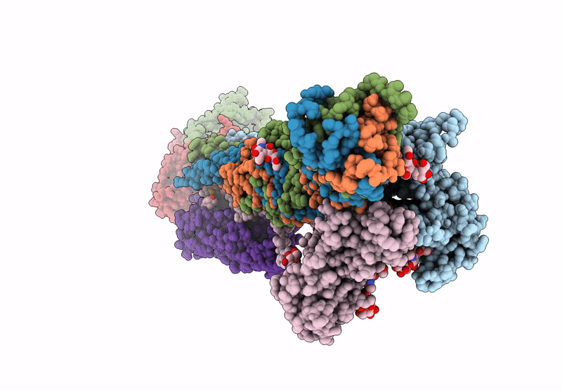
PDB ID:
8YT8
Keywords:
Title:
Cryo-EM structure of the dystrophin glycoprotein complex
Biological Source:
Source Organism(s):
Mus musculus (Taxon ID: 10090)
Method Details:
Experimental Method:
Resolution:
3.50 Å
Aggregation State:
PARTICLE
Reconstruction Method:
SINGLE PARTICLE