8YRV image
Entry Detail
PDB ID:
8YRV
Keywords:
Title:
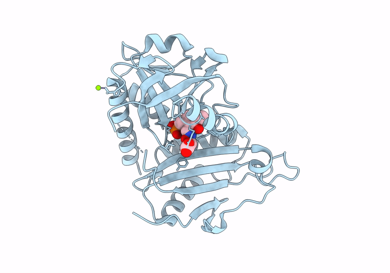
Crystal structure of D-amino acid transaminase from Haliscomenobacter hydrossis complexed with 3-aminooxypropionic acid
Biological Source:
Host Organism:
PDB Version:
Deposition Date:
2024-03-21
Release Date:
2024-04-17
Method Details:
Experimental Method:
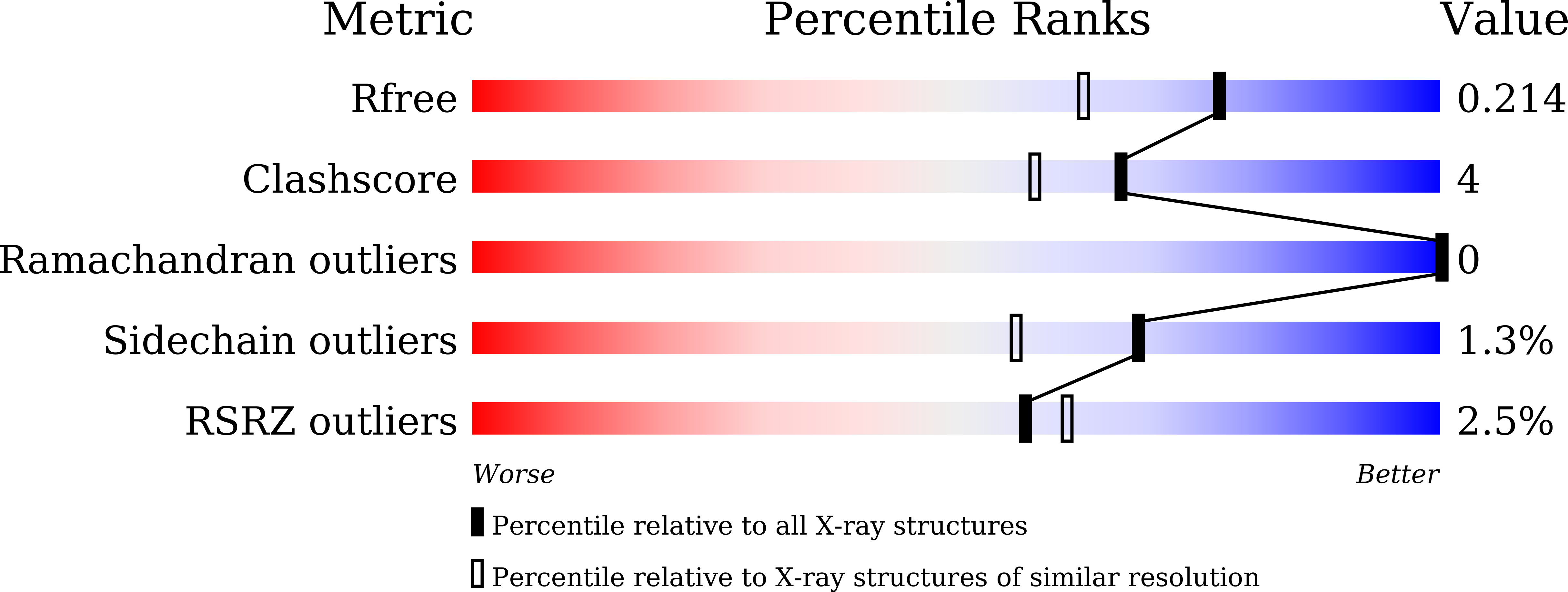
Resolution:
1.70 Å
R-Value Free:
0.21
R-Value Work:
0.16
R-Value Observed:
0.16
Space Group:
C 1 2 1
Macromolecular Entities
Polymer Type:polypeptide(L)
Description:Aminotransferase class IV
Chain IDs:A
Chain Length:283
Number of Molecules:1
Biological Source:Haliscomenobacter hydrossis DSM 1100
Primary Citation
Insights into the Functioning of the D-amino Acid Transaminase from Haliscomenobacter Hydrossis via a Structural and Spectral Analysis of its Complex with 3-Aminooxypropionic Acid.
Acta Naturae 16 18 24 (2024)
PMID: 39539521 DOI: 10.32607/actanaturae.27496

Abstact

Pyridoxal 5'-phosphate-dependent enzymes play a crucial role in nitrogen metabolism. Carbonyl compounds, such as O-substituted hydroxylamines, stand out among numerous specific inhibitors of these enzymes, including those of practical importance, because they react with pyridoxal 5'-phosphate in the active site of the enzymes to form stable oximes. O-substituted hydroxylamines mimic the side group of amino acid substrates, thus providing highly potent and specific inhibition of the corresponding enzymes. The interaction between D-amino acid transaminase from bacterium Haliscomenobacter hydrossis and 3-aminooxypropionic acid was studied in the present work. The structural and spectral analyses of the complex of this transaminase with 3-aminooxypropionic acid allowed us to clarify some features of the organization and functioning of its active site and illustrate one of the mechanisms of inhibition by the specific substrate, D-glutamic acid.

Legend

Protein

Chemical

Disease

Primary Citation of related structures