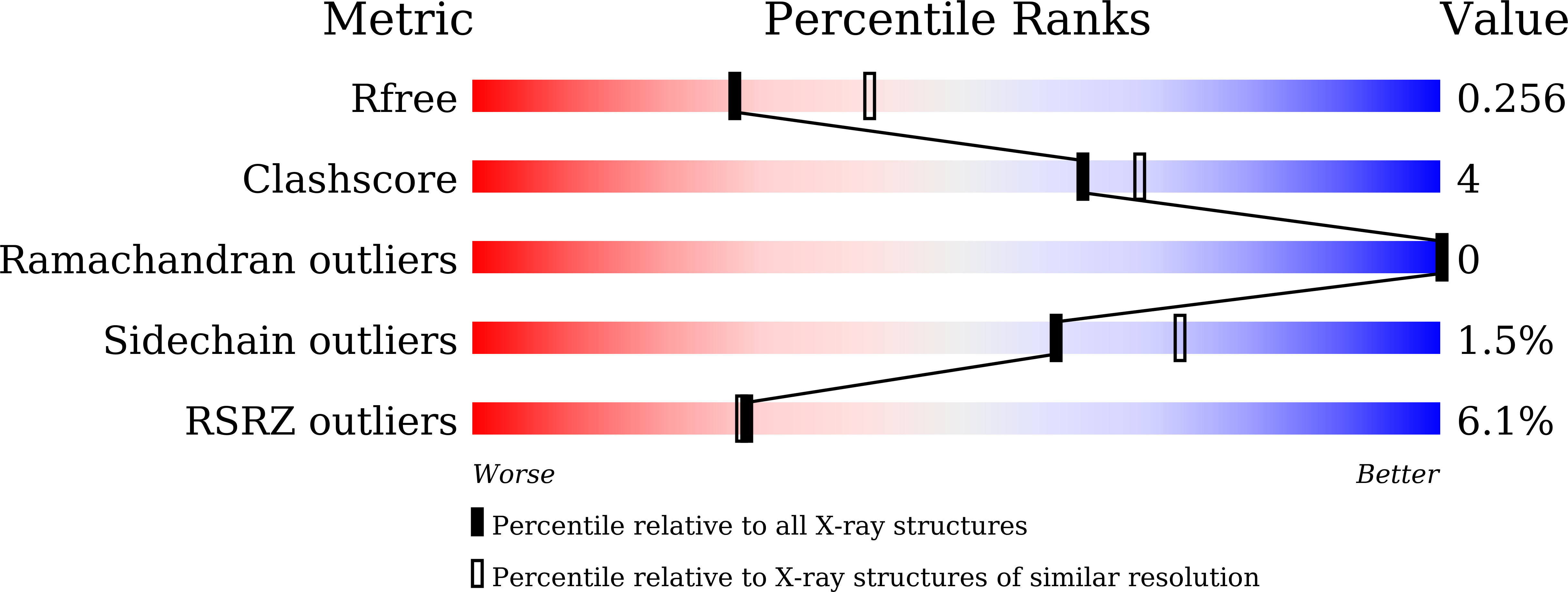
8YR6 image
Entry Detail
PDB ID:
8YR6
Keywords:
Title:
Crystal structure of E. coli phosphatidylserine synthase complexed with 16:0/16:0 CDP-DG
Biological Source:
PDB Version:
Deposition Date:
2024-03-20
Release Date:
2024-11-20
Method Details:
Experimental Method:
Resolution:
2.44 Å
R-Value Free:
0.24
R-Value Work:
0.21
R-Value Observed:
0.21
Space Group:
I 2 3
Macromolecular Entities
Polymer Type:polypeptide(L)
Description:CDP-diacylglycerol--serine O-phosphatidyltransferase
Mutations:H357A
Chain IDs:A
Chain Length:461
Number of Molecules:1
Biological Source:Escherichia coli str. K-12 substr. MG1655
Primary Citation
Structural basis for membrane association and catalysis by phosphatidylserine synthase in Escherichia coli.
Sci Adv 10 eadq4624 eadq4624 (2024)
PMID: 39693441 DOI: 10.1126/sciadv.adq4624

Abstact

Phosphatidylserine synthase (PssA) is essential in the biosynthesis of phosphatidylethanolamine, a major phospholipid of bacterial membranes. A peripheral membrane protein PssA can associate with the cellular membrane in its active state or exist in the cytosol in an inactive form. The membrane-bound enzyme acts on cytidine diphosphate diacylglycerol (CDP-DG) to form cytidine monophosphate and a covalent intermediate, which is subsequently targeted by serine to produce phosphatidylserine. Here, we present two crystal structures of Escherichia coli PssA, one complexed with CDP-DG and the other without. The lipid-bound structure mimics the Michaelis complex before the formation of a covalent intermediate, revealing key determinants for substrate recognition and catalysis. Notably, membrane-free PssA is in a monomer-dimer equilibrium, with only the monomer capable of associating with the membrane, suggesting a regulatory mechanism for phospholipid biosynthesis dependent on the oligomerization state of the enzyme.

Legend

Protein

Chemical

Disease

Primary Citation of related structures