8YNV image
Entry Detail
PDB ID:
8YNV
Keywords:
Title:
Poly(3-hydroxybutyrate) depolymerase PhaZ from Bacillus thuringiensis
Biological Source:
Source Organism:
Host Organism:
PDB Version:
Deposition Date:
2024-03-12
Release Date:
2024-12-11
Method Details:
Experimental Method:
Resolution:
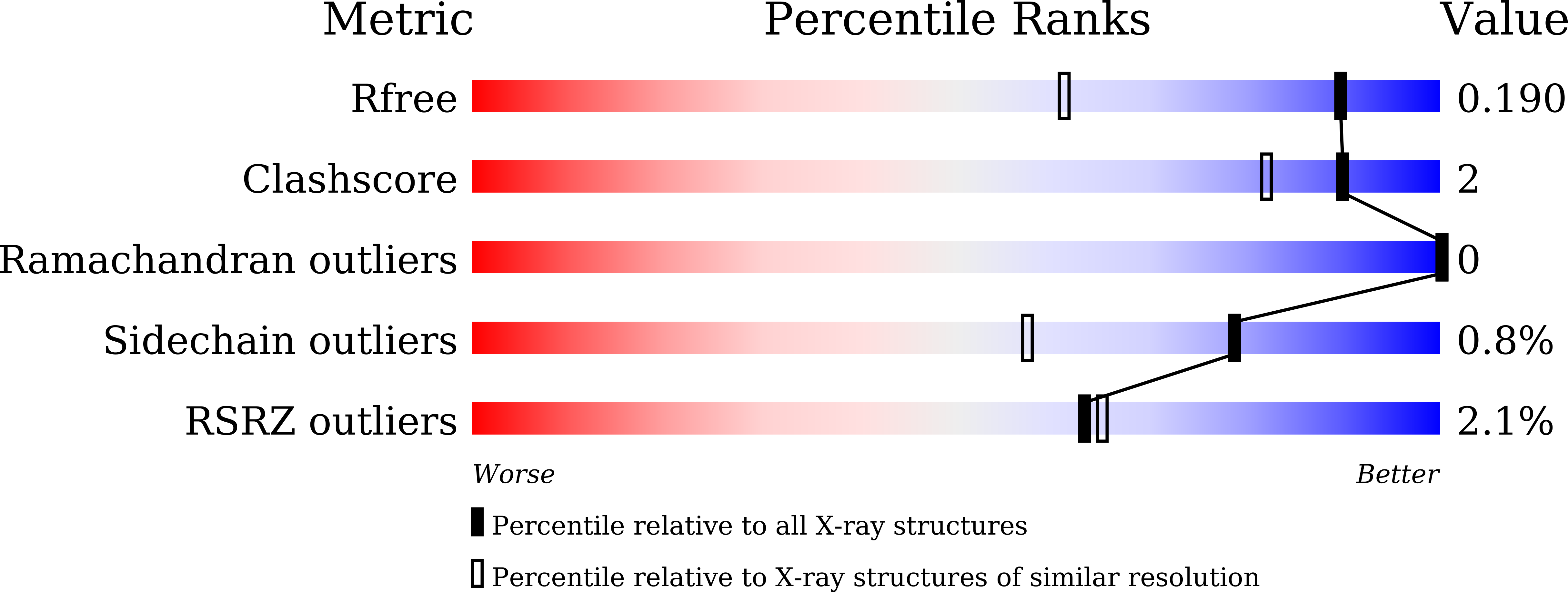
1.42 Å
R-Value Free:
0.18
R-Value Work:
0.16
R-Value Observed:
0.16
Space Group:
P 1
Macromolecular Entities
Polymer Type:polypeptide(L)
Description:poly(3-hydroxybutyrate) depolymerase
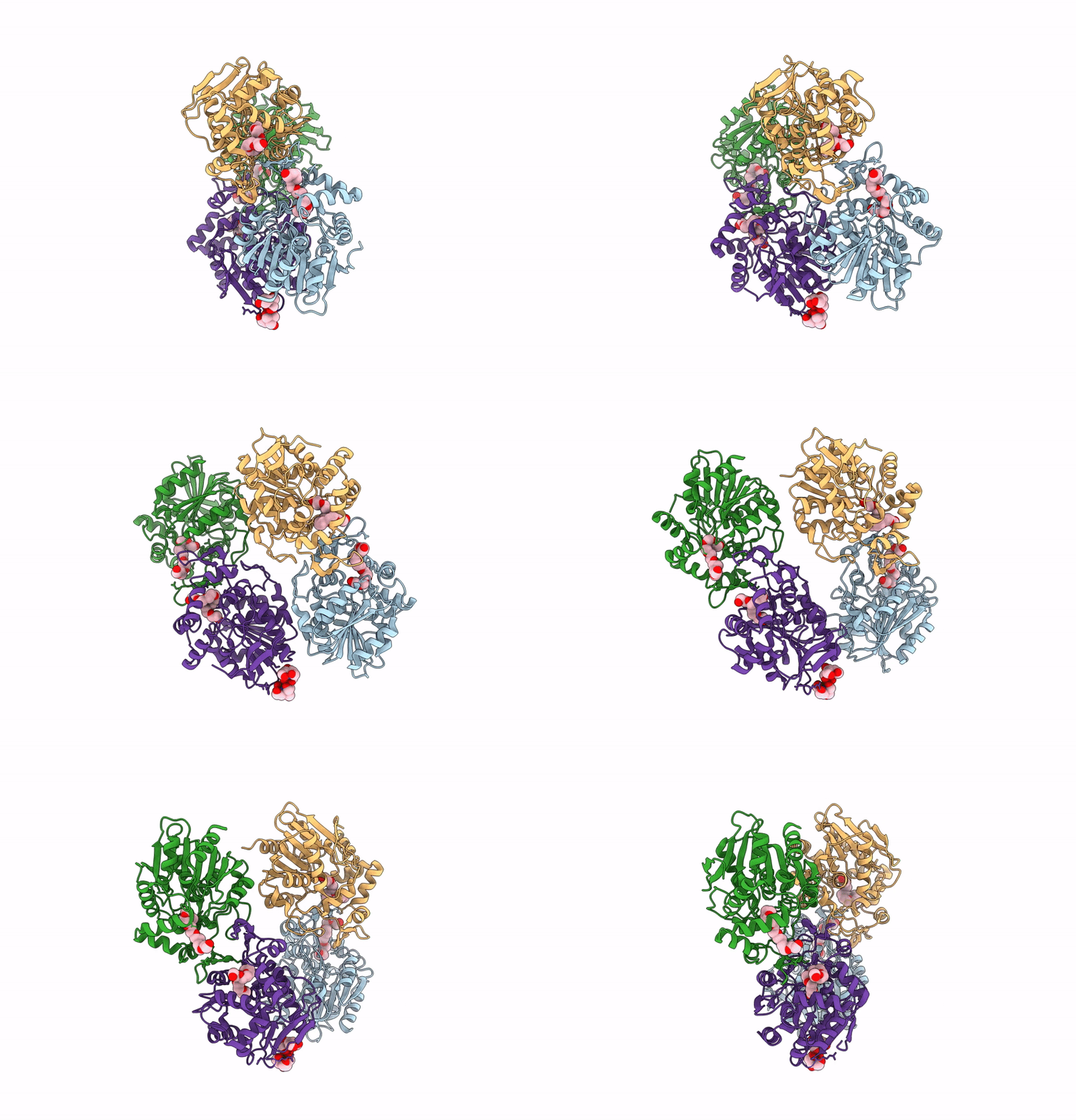
Chain IDs:A, B, C, D
Chain Length:311
Number of Molecules:4
Biological Source:Bacillus thuringiensis
Primary Citation
Structural insight into the poly(3-hydroxybutyrate) hydrolysis by intracellular PHB depolymerase from Bacillus thuringiensis.
Int.J.Biol.Macromol. 284 137999 137999 (2024)
PMID: 39592048 DOI: 10.1016/j.ijbiomac.2024.137999

Abstact

Poly((R)-3-hydroxybutyrate) (PHB) is a microbial biopolymer widely used in commercial biodegradable plastics. PHB degradation in cell is catalyzed by PHB depolymerase (PhaZ), which hydrolyzes the polyester into mono- and/or oligomeric (R)-3-hydroxylbutyrates (3HB). A novel intracellular PhaZ from Bacillus thuringiensis (BtPhaZ) was identified for potential applications in polymer biodegradation and 3HB production. Herein, we present the crystal structure of BtPhaZ at 1.42-Å resolution, making the first crystal structure for an intracellular PhaZ. BtPhaZ comprises a canonical α/β hydrolase catalytic domain and a unique α-helical cap domain. Despite lacking sequence similarity, BtPhaZ shares high structural homology with many α/β hydrolase members, exhibiting a similar active-site architecture. Alongside the most conserved superfamily signature, several new conserved signatures have been identified, contributing not only to the formations of the Ser-His-Asp catalytic triad and the oxyanion hole but also to the active-site conformation. The putative P-1 subsite appears to have limited space for accommodating only one 3HB-monomer, which may provide an explanation why the major hydrolytic product for BtPhaZ is monomeric form. Furthermore, a cluster of solvent-exposed hydrophobic residues in the helical cap domain forms an adsorption site for polymer-binding. Detailed structural comparisons reveal that various PhaZs employ distinct residues for the biopolymer-binding and hydrolysis.

Legend

Protein

Chemical

Disease

Primary Citation of related structures