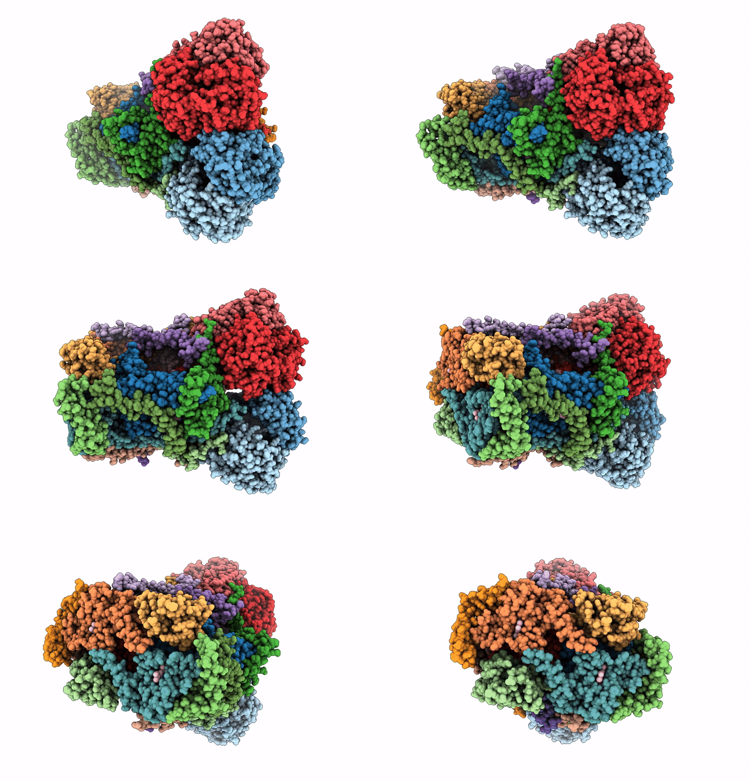
8YHQ image
Entry Detail
PDB ID:
8YHQ
EMDB ID:
Title:
Cryo-EM structure of Saccharomyces cerevisiae bc1 complex in pyraclostrobin-bound state
Biological Source:
Source Organism:
PDB Version:
Deposition Date:
2024-02-28
Release Date:
2024-12-25
Method Details:
Experimental Method:
Resolution:
2.42 Å
Aggregation State:
PARTICLE
Reconstruction Method:
SINGLE PARTICLE
Macromolecular Entities
Polymer Type:polypeptide(L)
Description:COR1 isoform 1
Chain IDs:A, K (auth: J)
Chain Length:431
Number of Molecules:2
Biological Source:Saccharomyces cerevisiae
Polymer Type:polypeptide(L)
Description:Cytochrome b-c1 complex subunit 2, mitochondrial
Chain IDs:C (auth: B), L (auth: K)
Chain Length:352
Number of Molecules:2
Biological Source:Saccharomyces cerevisiae
Polymer Type:polypeptide(L)
Description:Cytochrome b
Chain IDs:B (auth: C), G (auth: L)
Chain Length:385
Number of Molecules:2
Biological Source:Saccharomyces cerevisiae
Polymer Type:polypeptide(L)
Description:quinol--cytochrome-c reductase
Chain IDs:D, M
Chain Length:248
Number of Molecules:2
Biological Source:Saccharomyces cerevisiae
Polymer Type:polypeptide(L)
Description:Cytochrome b-c1 complex subunit Rieske, mitochondrial
Chain IDs:E, N
Chain Length:185
Number of Molecules:2
Biological Source:Saccharomyces cerevisiae
Polymer Type:polypeptide(L)
Description:QCR6 isoform 1
Chain IDs:F, P (auth: O)
Chain Length:75
Number of Molecules:2
Biological Source:Saccharomyces cerevisiae
Polymer Type:polypeptide(L)
Description:Cytochrome b-c1 complex subunit 7
Chain IDs:H (auth: G), Q (auth: P)
Chain Length:126
Number of Molecules:2
Biological Source:Saccharomyces cerevisiae
Polymer Type:polypeptide(L)
Description:Cytochrome b-c1 complex subunit 8
Chain IDs:I (auth: H), O (auth: Q)
Chain Length:93
Number of Molecules:2
Biological Source:Saccharomyces cerevisiae
Polymer Type:polypeptide(L)
Description:Cytochrome b-c1 complex subunit 9, mitochondrial
Chain IDs:J (auth: I), R
Chain Length:55
Number of Molecules:2
Biological Source:Saccharomyces cerevisiae
Polymer Type:polypeptide(L)
Description:Cytochrome b-c1 complex subunit 10, mitochondrial
Chain IDs:S, T
Chain Length:52
Number of Molecules:2
Biological Source:Saccharomyces cerevisiae
Primary Citation
Cryo-EM Structures Reveal the Unique Binding Modes of Metyltetraprole in Yeast and Porcine Cytochrome bc 1 Complex Enabling Rational Design of Inhibitors.
J.Am.Chem.Soc. 146 33903 33913 (2024)
PMID: 39601138 DOI: 10.1021/jacs.4c12595

Abstact

Cytochrome bc1 (complex III) represents a significant target for the discovery of both drugs and fungicides. Metyltetraprole (MET) is commonly classified as a quinone site inhibitor (QoI) that combats the G143A mutated isolate, which confers high resistance to strobilurin fungicides such as pyraclostrobin (PYR). The binding mode and antiresistance mechanism of MET remain unclear. Here, we determined the high-resolution structures of inhibitor-bound S. cerevisiae complex III (MET, 2.52 Å; PYR, 2.42 Å) and inhibitor-bound porcine complex III (MET, 2.53 Å; PYR, 2,37 Å) by cryo-electron microscopy. The distinct binding modes of MET and PYR were observed for the first time. Notably, the MET exhibited different binding modes in the two species. In S. cerevisiae, the binding site of MET was the same as PYR, serving as a Pm-type inhibitor of the Qo site. However, in porcine, MET acted as a dual-target inhibitor of both Qo and Qi. Based on the structural insights, a novel inhibitor (YF23694) was discovered and demonstrated excellent fungicidal activity against downy mildew and powdery mildew fungi. This work provides a new starting point for the design of the next generation of inhibitors to overcome the resistance.

Legend

Protein

Chemical

Disease

Primary Citation of related structures