Deposition Date
2024-02-26
Release Date
2024-09-11
Last Version Date
2024-10-30
Entry Detail
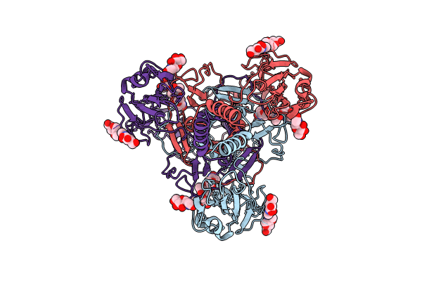
PDB ID:
8YG6
Keywords:
Title:
The pre-fusion structure of baculovirus fusion protein GP64
Biological Source:
Source Organism(s):
Autographa californica nucleopolyhedrovirus (Taxon ID: 46015)
Expression System(s):
Method Details:
Experimental Method:
Resolution:
2.77 Å
Aggregation State:
PARTICLE
Reconstruction Method:
SINGLE PARTICLE