8Y9R image
Entry Detail
PDB ID:
8Y9R
Keywords:
Title:
Crystal structure of Spiral2 microtubule-binding domain from Physcomitrella patens
Biological Source:
Source Organism:
PDB Version:
Deposition Date:
2024-02-07
Release Date:
2024-12-18
Method Details:
Experimental Method:
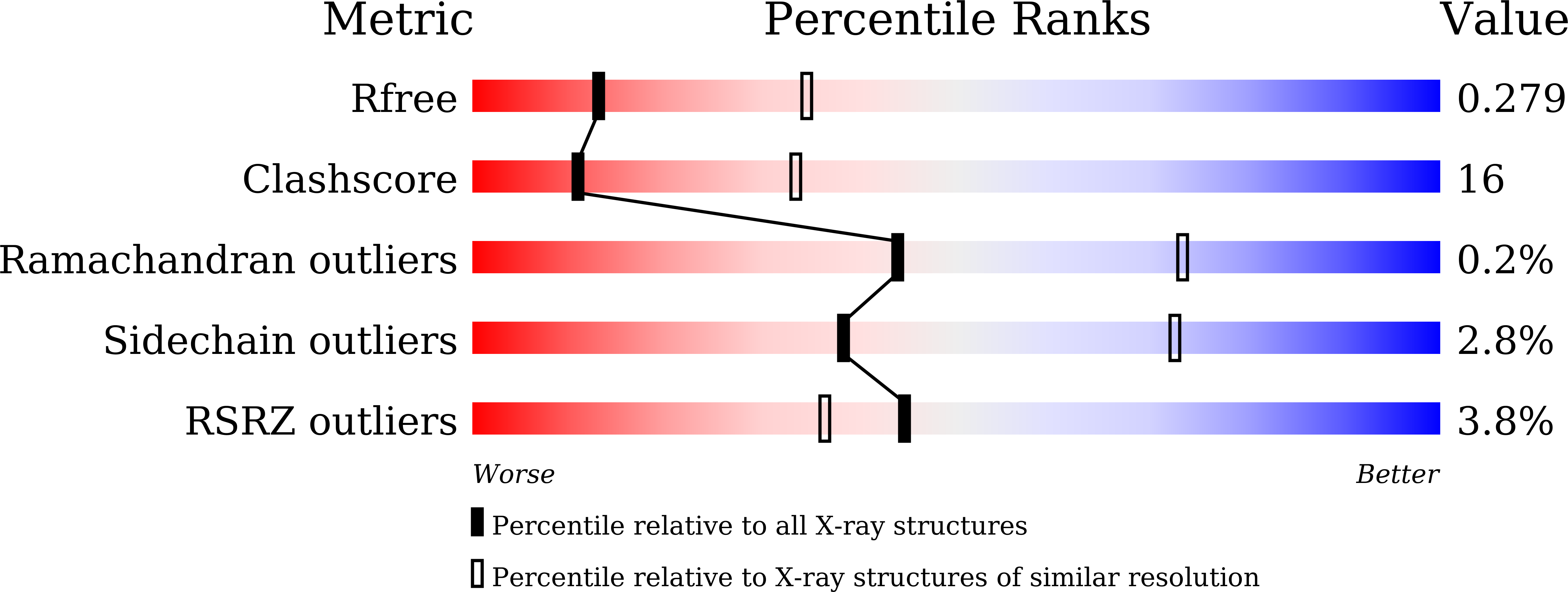
Resolution:
2.80 Å
R-Value Free:
0.27
R-Value Work:
0.21
Space Group:
P 2 21 21
Macromolecular Entities
Polymer Type:polypeptide(L)
Description:Microtubule binding protein
Chain IDs:A, B
Chain Length:287
Number of Molecules:2
Biological Source:Physcomitrium patens
Modified Residue
Compound ID Chain ID Parent Comp ID Details 2D Image
MSE A MET modified residue
Primary Citation
Structural analysis of microtubule binding by minus-end targeting protein Spiral2.
Biochim Biophys Acta Mol Cell Res 1871 119858 119858 (2024)
PMID: 39370045 DOI: 10.1016/j.bbamcr.2024.119858

Abstact

Microtubules (MTs) are dynamic cytoskeletal polymers that play a critical role in determining cell polarity and shape. In plant cells, acentrosomal MTs are localized on the cell surface and are referred to as cortical MTs. Cortical MTs nucleate in the cell cortex and detach from nucleation sites. The released MT filaments perform treadmilling, with the plus-ends of MTs polymerizing and the minus-ends depolymerizing. Minus-end targeting proteins, -TIPs, include Spiral2, which regulates the minus-end dynamics of acentrosomal MTs. Spiral2 accumulates autonomously at MT minus-ends and inhibits filament shrinkage, but the mechanism by which Spiral2 specifically recognizes minus-ends of MTs remains unknown. Here we describe the crystal structure of Spiral2's N-terminal MT-binding domain. The structural properties of this domain resemble those of the HEAT repeat structure of the tumor overexpressed gene (TOG) domain, but the number of HEAT repeats is different and the conformation is highly arched. Gel filtration and co-sedimentation analyses demonstrate that the domain binds preferentially to MT filaments rather than the tubulin dimer, and that the tubulin-binding mode of Spiral2 via the basic surface is similar to that of the TOG domain. We constructed an in silico model of the Spiral2-tubulin complex to identify residues that potentially recognize tubulin. Mutational analysis revealed that the key residues inferred in the model are involved in microtubule recognition, and provide insight into the mechanism by which end-targeting proteins stabilize MT ends.

Legend

Protein

Chemical

Disease

Primary Citation of related structures