Deposition Date
2024-01-19
Release Date
2024-11-13
Last Version Date
2024-11-27
Entry Detail
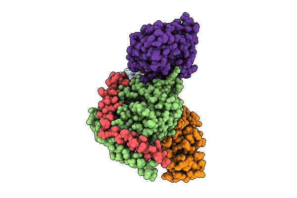
PDB ID:
8XYD
Keywords:
Title:
Structure of Platelet-activating factor receptor-G protein complex bound to platelet-activating factor
Biological Source:
Source Organism(s):
Homo sapiens (Taxon ID: 9606)
Mus musculus (Taxon ID: 10090)
Mus musculus (Taxon ID: 10090)
Expression System(s):
Method Details:
Experimental Method:
Resolution:
2.90 Å
Aggregation State:
PARTICLE
Reconstruction Method:
SINGLE PARTICLE