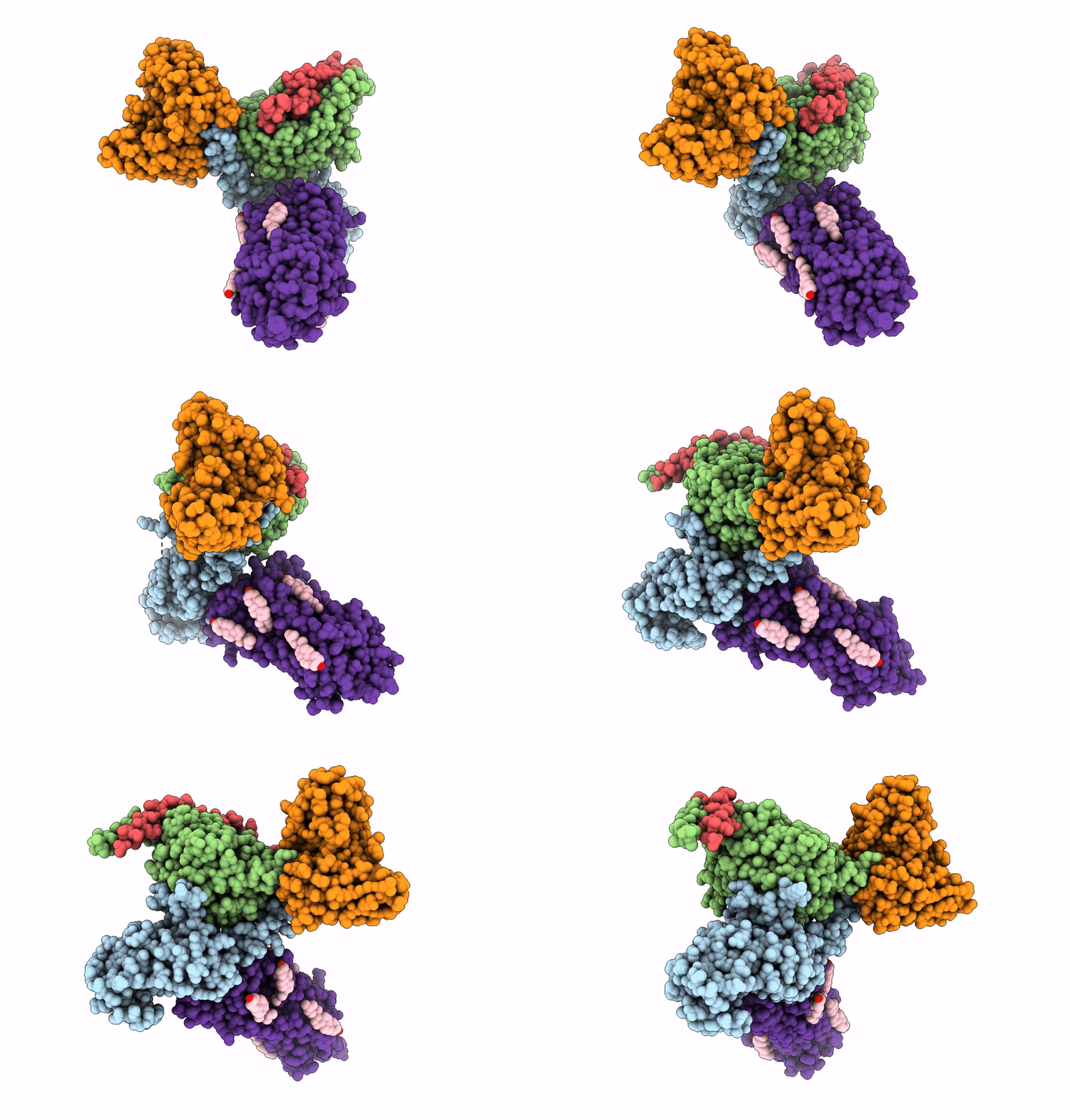
8XOR image
Entry Detail
PDB ID:
8XOR
EMDB ID:
Title:
Cryo-EM structure of the tethered agonist-bound human PAR1-Gq complex
Biological Source:
Host Organism:
PDB Version:
Deposition Date:
2024-01-02
Release Date:
2024-09-18
Method Details:
Experimental Method:
Resolution:
3.00 Å
Aggregation State:
PARTICLE
Reconstruction Method:
SINGLE PARTICLE
Macromolecular Entities
Polymer Type:polypeptide(L)
Description:G subunit q (Gi1-Gq chimeric)
Chain IDs:C (auth: A)
Chain Length:361
Number of Molecules:1
Biological Source:Homo sapiens
Polymer Type:polypeptide(L)
Description:Guanine nucleotide-binding protein G(I)/G(S)/G(T) subunit beta-1
Chain IDs:A (auth: B)
Chain Length:379
Number of Molecules:1
Biological Source:Rattus
Polymer Type:polypeptide(L)
Description:Guanine nucleotide-binding protein G(I)/G(S)/G(O) subunit gamma-2
Chain IDs:B (auth: C)
Chain Length:67
Number of Molecules:1
Biological Source:Bos taurus
Polymer Type:polypeptide(L)
Description:scFv16
Chain IDs:D (auth: E)
Chain Length:303
Number of Molecules:1
Biological Source:Mus musculus
Polymer Type:polypeptide(L)
Description:Proteinase-activated receptor 1 LgBiT
Chain IDs:E (auth: R)
Chain Length:532
Number of Molecules:1
Biological Source:Homo sapiens
Ligand Molecules
Primary Citation
Structural basis of tethered agonism and G protein coupling of protease-activated receptors.
Cell Res. 34 725 734 (2024)
PMID: 38997424 DOI: 10.1038/s41422-024-00997-2

Abstact

Protease-activated receptors (PARs) are a unique group within the G protein-coupled receptor superfamily, orchestrating cellular responses to extracellular proteases via enzymatic cleavage, which triggers intracellular signaling pathways. Protease-activated receptor 1 (PAR1) is a key member of this family and is recognized as a critical pharmacological target for managing thrombotic disorders. In this study, we present cryo-electron microscopy structures of PAR1 in its activated state, induced by its natural tethered agonist (TA), in complex with two distinct downstream proteins, the Gq and Gi heterotrimers, respectively. The TA peptide is positioned within a surface pocket, prompting PAR1 activation through notable conformational shifts. Contrary to the typical receptor activation that involves the outward movement of transmembrane helix 6 (TM6), PAR1 activation is characterized by the simultaneous downward shift of TM6 and TM7, coupled with the rotation of a group of aromatic residues. This results in the displacement of an intracellular anion, creating space for downstream G protein binding. Our findings delineate the TA recognition pattern and highlight a distinct role of the second extracellular loop in forming β-sheets with TA within the PAR family, a feature not observed in other TA-activated receptors. Moreover, the nuanced differences in the interactions between intracellular loops 2/3 and the Gα subunit of different G proteins are crucial for determining the specificity of G protein coupling. These insights contribute to our understanding of the ligand binding and activation mechanisms of PARs, illuminating the basis for PAR1's versatility in G protein coupling.

Legend

Protein

Chemical

Disease

Primary Citation of related structures