8XJV image
Entry Detail
PDB ID:
8XJV
EMDB ID:
Keywords:
Title:
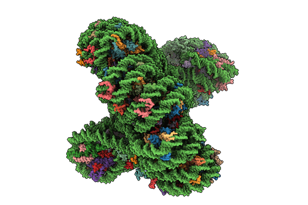
Structural basis for the linker histone H5-nucleosome binding and chromatin compaction
Biological Source:
PDB Version:
Deposition Date:
2023-12-22
Release Date:
2024-09-11
Method Details:
Experimental Method:
Resolution:
3.60 Å
Aggregation State:
PARTICLE
Reconstruction Method:
SINGLE PARTICLE
Macromolecular Entities
Polymer Type:polypeptide(L)
Description:Histone H2A
Chain IDs:C (auth: A), D (auth: B), E (auth: C), F (auth: D), G (auth: E), H (auth: F), I (auth: G), J (auth: H), K (auth: I), M (auth: K), N (auth: L), VB (auth: aj), WB (auth: ak), XB (auth: al), YB (auth: am), ZB (auth: an), AC (auth: ao), BC (auth: ap), CC (auth: aq), DC (auth: ar), EC (auth: as), FC (auth: at), GC (auth: au), QC (auth: Ae)
Chain Length:130
Number of Molecules:24
Biological Source:Xenopus laevis
Polymer Type:polypeptide(L)
Description:Histone H5
Chain IDs:TC (auth: Ah), UC (auth: Ai), VC (auth: Aj), WC (auth: Ak), XC (auth: Al), YC (auth: Am), ZC (auth: An), AD (auth: Ao), BD (auth: Ap), CD (auth: Aq), DD (auth: Ar), ED (auth: As)
Chain Length:196
Number of Molecules:12
Biological Source:Gallus gallus
Polymer Type:polydeoxyribonucleotide
Description:DNA
Chain IDs:A (auth: Au)
Chain Length:2124
Number of Molecules:1
Biological Source:synthetic construct
Polymer Type:polydeoxyribonucleotide
Description:DNA
Chain IDs:B (auth: Av)
Chain Length:123
Number of Molecules:1
Biological Source:synthetic construct
Polymer Type:polypeptide(L)
Description:Histone H2B 1.1
Chain IDs:L (auth: J), O (auth: M), P (auth: N), Q (auth: O), R (auth: P), S (auth: Q), T (auth: R), U (auth: S), V (auth: T), W (auth: U), X (auth: V), Y (auth: W), HC (auth: av), IC (auth: aw), JC (auth: ax), KC (auth: ay), LC (auth: az), MC (auth: Aa), NC (auth: Ab), OC (auth: Ac), PC (auth: Ad), RC (auth: Af), SC (auth: Ag), FD (auth: At)
Chain Length:123
Number of Molecules:24
Biological Source:Xenopus laevis
Polymer Type:polypeptide(L)
Description:Histone H3
Chain IDs:Z (auth: X), AA (auth: Y), BA (auth: Z), CA (auth: a), DA (auth: b), EA (auth: c), FA (auth: d), GA (auth: e), HA (auth: f), IA (auth: g), JA (auth: h), KA (auth: i), LA (auth: j), MA (auth: k), NA (auth: l), OA (auth: m), PA (auth: n), QA (auth: o), RA (auth: p), SA (auth: q), TA (auth: r), UA (auth: s), VA (auth: t), WA (auth: u)
Chain Length:130
Number of Molecules:24
Biological Source:Xenopus laevis
Polymer Type:polypeptide(L)
Description:Histone H4
Chain IDs:XA (auth: v), YA (auth: w), ZA (auth: x), AB (auth: y), BB (auth: z), CB (auth: 0), DB (auth: 1), EB (auth: 2), FB (auth: 3), GB (auth: 4), HB (auth: 5), IB (auth: 6), JB (auth: 7), KB (auth: 8), LB (auth: 9), MB (auth: CD), NB (auth: a2), OB (auth: a3), PB (auth: a4), QB (auth: a5), RB (auth: a6), SB (auth: a7), TB (auth: a8), UB (auth: a9)
Chain Length:103
Number of Molecules:24
Biological Source:Xenopus laevis
Ligand Molecules
Primary Citation
Structural basis for linker histone H5-nucleosome binding and chromatin fiber compaction.
Cell Res. 34 707 724 (2024)
PMID: 39103524 DOI: 10.1038/s41422-024-01009-z

Abstact

The hierarchical packaging of chromatin fibers plays a critical role in gene regulation. The 30-nm chromatin fibers, a central-level structure bridging nucleosomal arrays to higher-order organizations, function as the first level of transcriptional dormant chromatin. The dynamics of 30-nm chromatin fiber play a crucial role in biological processes related to DNA. Here, we report a 3.6-angstrom resolution cryogenic electron microscopy structure of H5-bound dodecanucleosome, i.e., the chromatin fiber reconstituted in the presence of linker histone H5, which shows a two-start left-handed double helical structure twisted by tetranucleosomal units. An atomic structural model of the H5-bound chromatin fiber, including an intact chromatosome, is built, which provides structural details of the full-length linker histone H5, including its N-terminal domain and an HMG-motif-like C-terminal domain. The chromatosome structure shows that H5 binds the nucleosome off-dyad through a three-contact mode in the chromatin fiber. More importantly, the H5-chromatin structure provides a fine molecular basis for the intra-tetranucleosomal and inter-tetranucleosomal interactions. In addition, we systematically validated the physiological functions and structural characteristics of the tetranucleosomal unit through a series of genetic and genomic studies in Saccharomyces cerevisiae and in vitro biophysical experiments. Furthermore, our structure reveals that multiple structural asymmetries of histone tails confer a polarity to the chromatin fiber. These findings provide structural and mechanistic insights into how a nucleosomal array folds into a higher-order chromatin fiber with a polarity in vitro and in vivo.

Legend

Protein

Chemical

Disease

Primary Citation of related structures