8XJ3 image
Deposition Date 2023-12-20
Release Date 2024-06-05
Last Version Date 2024-06-12
Entry Detail
PDB ID:
8XJ3
Keywords:
Title:
Crystal structure of methyltransferase CbiL from Akkermansia muciniphila
Biological Source:
Source Organism:
Method Details:
Experimental Method:
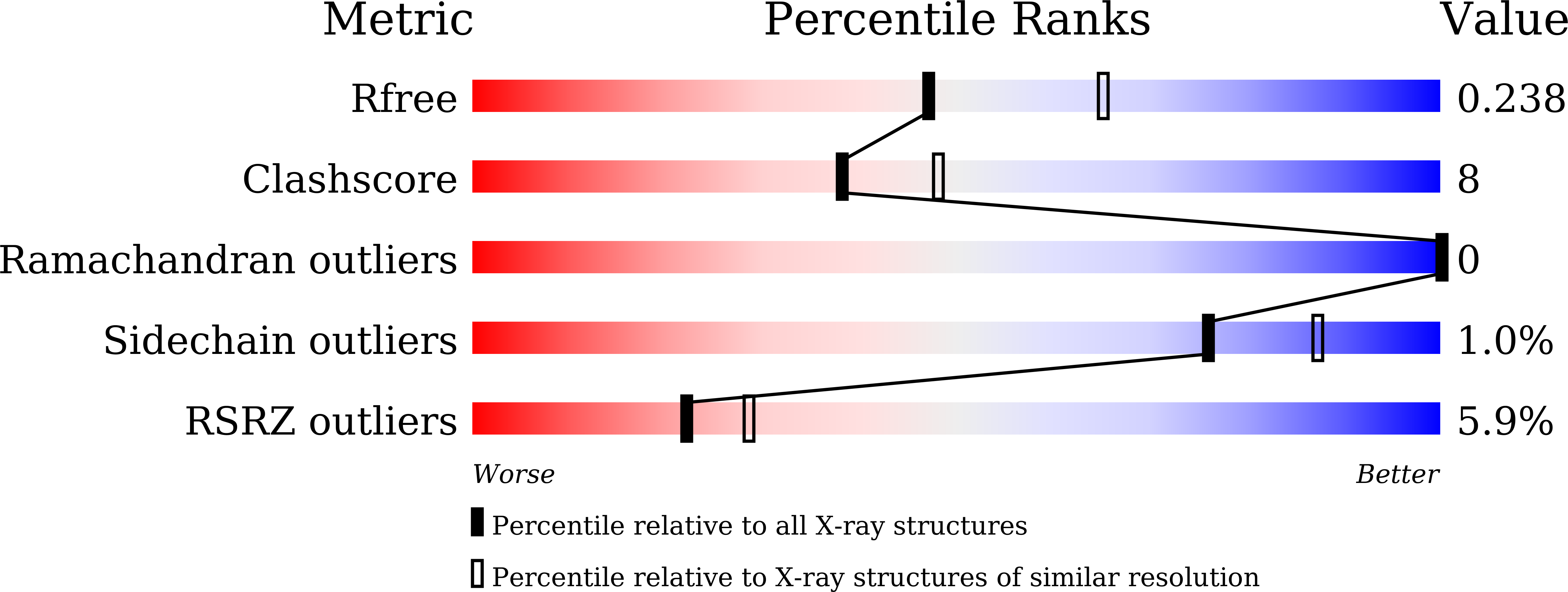
Resolution:
2.30 Å
R-Value Free:
0.24
R-Value Work:
0.19
R-Value Observed:
0.19
Space Group:
P 21 21 21
Macromolecular Entities
Polymer Type:polypeptide(L)
Molecule:CbiL
Chain IDs:A, B
Chain Length:255
Number of Molecules:2
Biological Source:Akkermansia muciniphila
Primary Citation
Crystal structure of methyltransferase CbiL from Akkermansia muciniphila.
Biochem.Biophys.Res.Commun. 722 150165 150165 (2024)
PMID: 38805786 DOI: 10.1016/j.bbrc.2024.150165

Abstact

Akkermansia muciniphila is a mucin-degrading probiotic that colonizes the gastrointestinal tract. Genomic analysis identified a set of genes involved in the biosynthesis of corrin ring, including the cobalt factor II methyltransferase CbiL, in some phylogroups of A. muciniphila, implying a potential capacity for de novo synthesis of cobalamin. In this work, we determined the crystal structure of CbiL from A. muciniphila at 2.3 Å resolution. AmCbiL exists as a dimer both in solution and in crystal, and each protomer consists of two α/β domains, the N-terminal domain and the C-terminal domain, consistent with the folding of typical class III MTases. The two domains create an open trough, potentially available to bind the substrates SAM and cobalt factor II. Sequence and structural comparisons with other CbiLs, assisted by computer modeling, suggest that AmCbiL should have cobalt factor II C-20 methyltransferase activity. Our results support that certain strains of A. muciniphila may be capable of synthesizing cobalamin de novo.

Legend

Protein

Chemical

Disease

Primary Citation of related structures