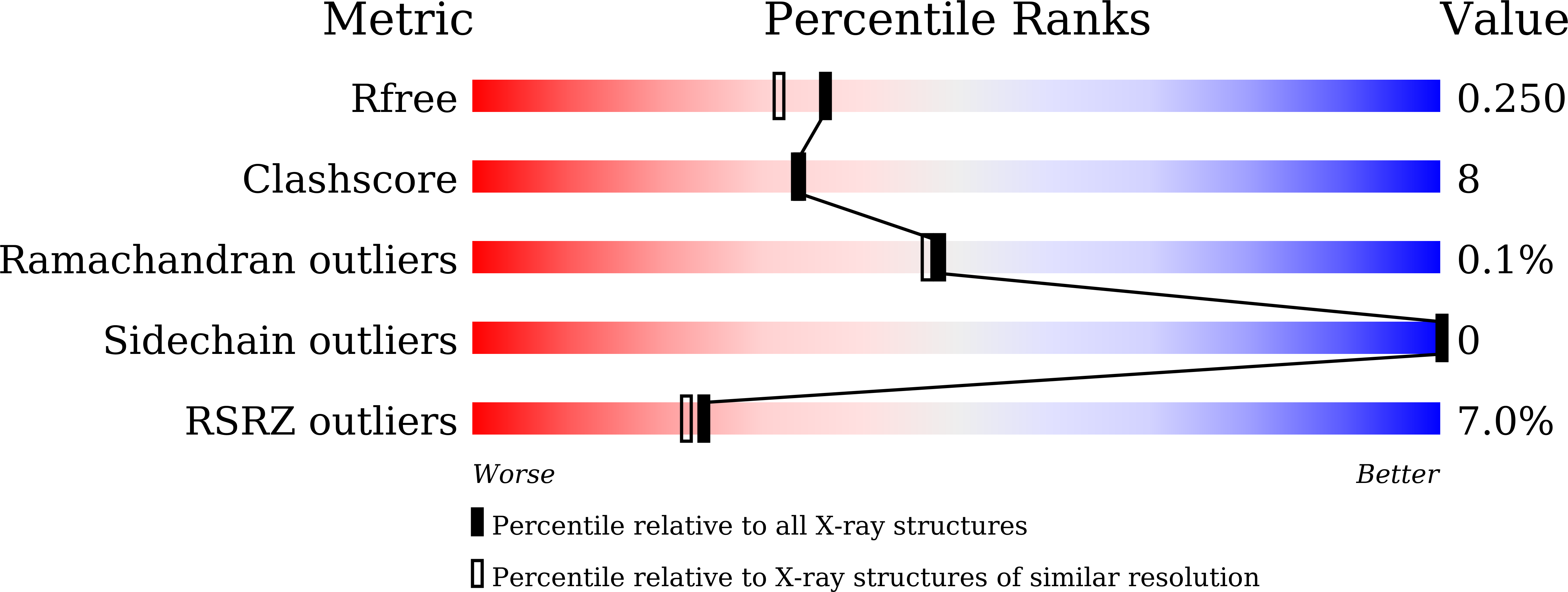
8X89 image
Deposition Date 2023-11-27
Release Date 2024-10-16
Last Version Date 2024-10-30
Entry Detail
PDB ID:
8X89
Keywords:
Title:
Crystal Structure of Streptococcus pneumoniae fabG
Biological Source:
Source Organism:
Method Details:
Experimental Method:
Resolution:
1.99 Å
R-Value Free:
0.24
R-Value Work:
0.21
R-Value Observed:
0.21
Space Group:
P 21 21 21
Macromolecular Entities
Structures with similar UniProt ID
Protein Blast
Polymer Type:polypeptide(L)
Molecule:3-oxoacyl-[acyl-carrier-protein] reductase
Chain IDs:A, B, C, D
Chain Length:243
Number of Molecules:4
Biological Source:Streptococcus pneumoniae
Primary Citation
Structure features of Streptococcus pneumoniae FabG and virtual screening of allosteric inhibitors.
Front Mol Biosci 11 1472252 1472252 (2024)
PMID: 39398278 DOI: 10.3389/fmolb.2024.1472252

Abstact

Streptococcus pneumoniae, a gram-positive bacterium, is responsible for diverse infections globally, and its antibiotic resistance presents significant challenges to medical advancements. It is imperative to employ various strategies to identify antibiotics. 3-oxoacyl-[acyl-carrier-protein] reductase (FabG) is a key component in the type II fatty acid synthase (FAS II) system, which is a developing target for new anti-streptococcal drugs. We first demonstrated the function of SpFabG in vivo and in vitro and the 2 Å SpFabG structure was elucidated using X-ray diffraction technique. It was observed that the NADPH binding promotes the transformation from tetramers to dimers in solution, suggesting dimers but not tetramer may be the active conformation. By comparing the structures of FabG homologues, we have identified the conserved tetramerization site and further confirmed the mechanism that the tetramerization site mutation leads to a loss of function and destabilization through mutagenesis experiments. Starting from 533,600 compounds, we proceeded with a sequential workflow involving pharmacophore-based virtual screening, molecular docking, and binding energy calculations. Combining all the structural analysis, we identified L1, L2 and L5 as a promising candidate for SpFabG inhibitor, based on the most stable binding mode in comparison to other evaluated inhibitors.

Legend

Protein

Chemical

Disease

Primary Citation of related structures