8X47 image
Entry Detail
PDB ID:
8X47
Keywords:
Title:
Crystal structure of DIMT1 in complex with S-adenosyl-L-homocysteine (SAH) from Pyrococcus horikoshii
Biological Source:
Source Organism:
PDB Version:
Deposition Date:
2023-11-15
Release Date:
2024-08-28
Method Details:
Experimental Method:
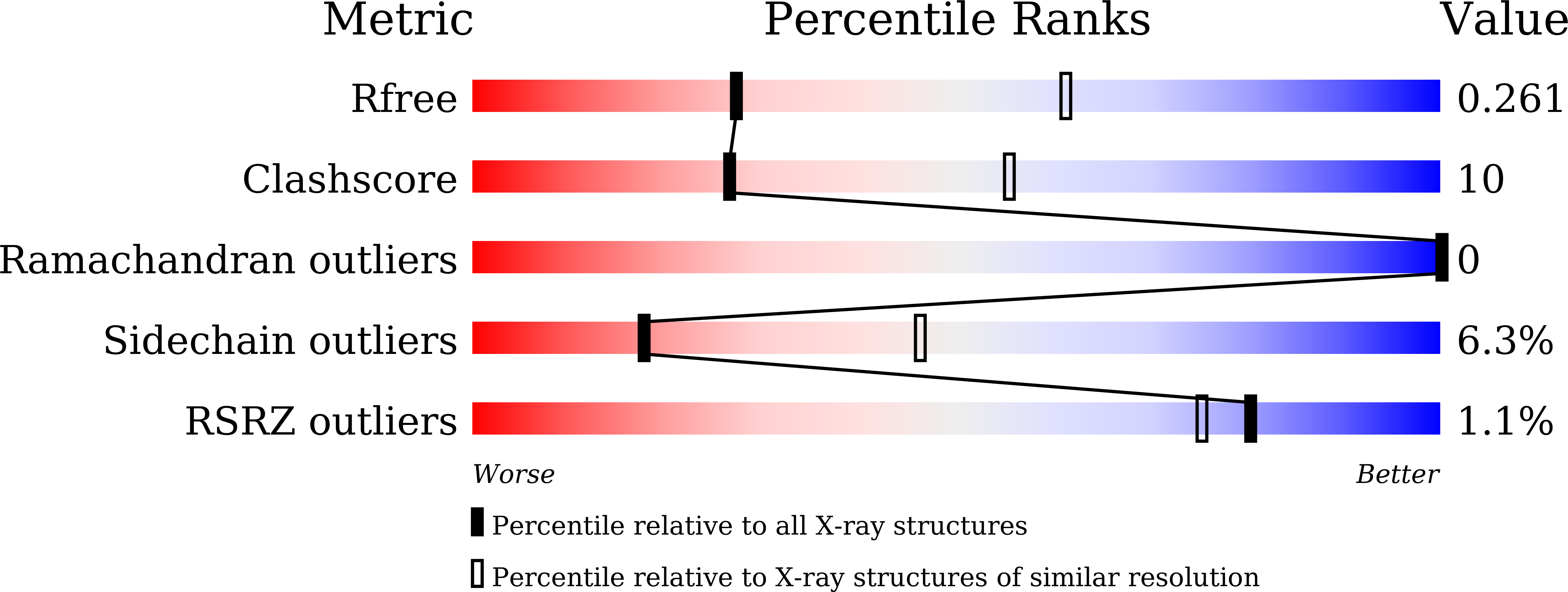
Resolution:
2.80 Å
R-Value Free:
0.27
R-Value Work:
0.18
R-Value Observed:
0.18
Space Group:
C 2 2 21
Macromolecular Entities
Polymer Type:polypeptide(L)
Description:Probable ribosomal RNA small subunit methyltransferase A
Chain IDs:A
Chain Length:290
Number of Molecules:1
Biological Source:Pyrococcus horikoshii OT3
Primary Citation
Structural and functional characterization of archaeal DIMT1 unveils distinct protein dynamics essential for efficient catalysis.
Structure 32 1760 1775.e7 (2024)
PMID: 39146930 DOI: 10.1016/j.str.2024.07.013

Abstact

Dimethyladenosine transferase 1 (DIMT1), an ortholog of bacterial KsgA is a conserved protein that assists in ribosome biogenesis by modifying two successive adenosine bases near the 3' end of small subunit (SSU) rRNA. Although KsgA/DIMT1 proteins have been characterized in bacteria and eukaryotes, they are yet unexplored in archaea. Also, their dynamics are not well understood. Here, we structurally and functionally characterized the apo and holo forms of archaeal DIMT1 from Pyrococcus horikoshii. Wild-type protein and mutants were analyzed to capture different transition states, including open, closed, and intermediate states. This study reports a unique inter-domain movement that is needed for substrate (RNA) positioning in the catalytic pocket, and is only observed in the presence of the cognate cofactors S-adenosyl-L-methionine (SAM) or S-adenosyl-L-homocysteine (SAH). The binding of the inhibitor sinefungine, an analog of SAM or SAH, to archaeal DIMT1 blocks the catalytic pocket and renders the enzyme inactive.

Legend

Protein

Chemical

Disease

Primary Citation of related structures