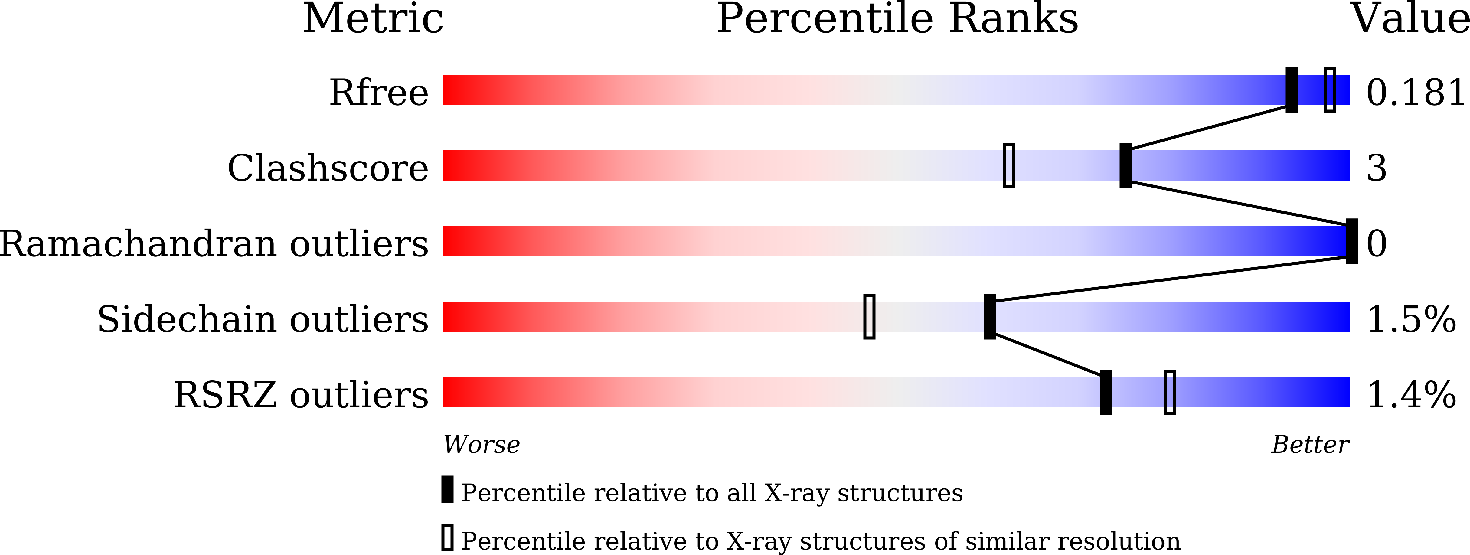
8X3G image
Deposition Date 2023-11-13
Release Date 2024-09-11
Last Version Date 2024-09-11
Entry Detail
PDB ID:
8X3G
Keywords:
Title:
Crystal structure of metformin hydrolase from Aminobacter
Biological Source:
Source Organism:
Method Details:
Experimental Method:
Resolution:
1.84 Å
R-Value Free:
0.18
R-Value Work:
0.15
R-Value Observed:
0.15
Space Group:
P 21 21 21
Macromolecular Entities
Polymer Type:polypeptide(L)
Molecule:Agmatinase family protein
Chain IDs:A, B
Chain Length:378
Number of Molecules:2
Biological Source:Aminobacter sp. NyZ550
Polymer Type:polypeptide(L)
Molecule:Arginase family protein
Chain IDs:C, D, E, F
Chain Length:348
Number of Molecules:4
Biological Source:Aminobacter sp. NyZ550
Primary Citation
Discovery of a Ni 2+ -dependent heterohexameric metformin hydrolase.
Nat Commun 15 6121 6121 (2024)
PMID: 39033196 DOI: 10.1038/s41467-024-50409-7

Abstact

The biguanide drug metformin is a first-line blood glucose-lowering medication for type 2 diabetes, leading to its presence in the global environment. However, little is known about the fate of metformin by microbial catabolism. Here, we characterize a Ni2+-dependent heterohexameric enzyme (MetCaCb) from the ureohydrolase superfamily, catalyzing the hydrolysis of metformin into guanylurea and dimethylamine. Either subunit alone is catalytically inactive, but together they work as an active enzyme highly specific for metformin. The crystal structure of the MetCaCb complex shows the coordination of the binuclear metal cluster only in MetCa, with MetCb as a protein binder of its active cognate. An in-silico search and functional assay discover a group of MetCaCb-like protein pairs exhibiting metformin hydrolase activity in the environment. Our findings not only establish the genetic and biochemical foundation for metformin catabolism but also provide additional insights into the adaption of the ancient enzymes toward newly occurred substrate.

Legend

Protein

Chemical

Disease

Primary Citation of related structures