Deposition Date
2023-10-30
Release Date
2024-06-26
Last Version Date
2024-07-17
Entry Detail
PDB ID:
8WXR
Keywords:
Title:
Structure of WDR5 in complex with WIN motif containing MBD3C F47A
Biological Source:
Source Organism(s):
Homo sapiens (Taxon ID: 9606)
Mus musculus (Taxon ID: 10090)
Mus musculus (Taxon ID: 10090)
Expression System(s):
Method Details:
Experimental Method:
Resolution:
2.08 Å
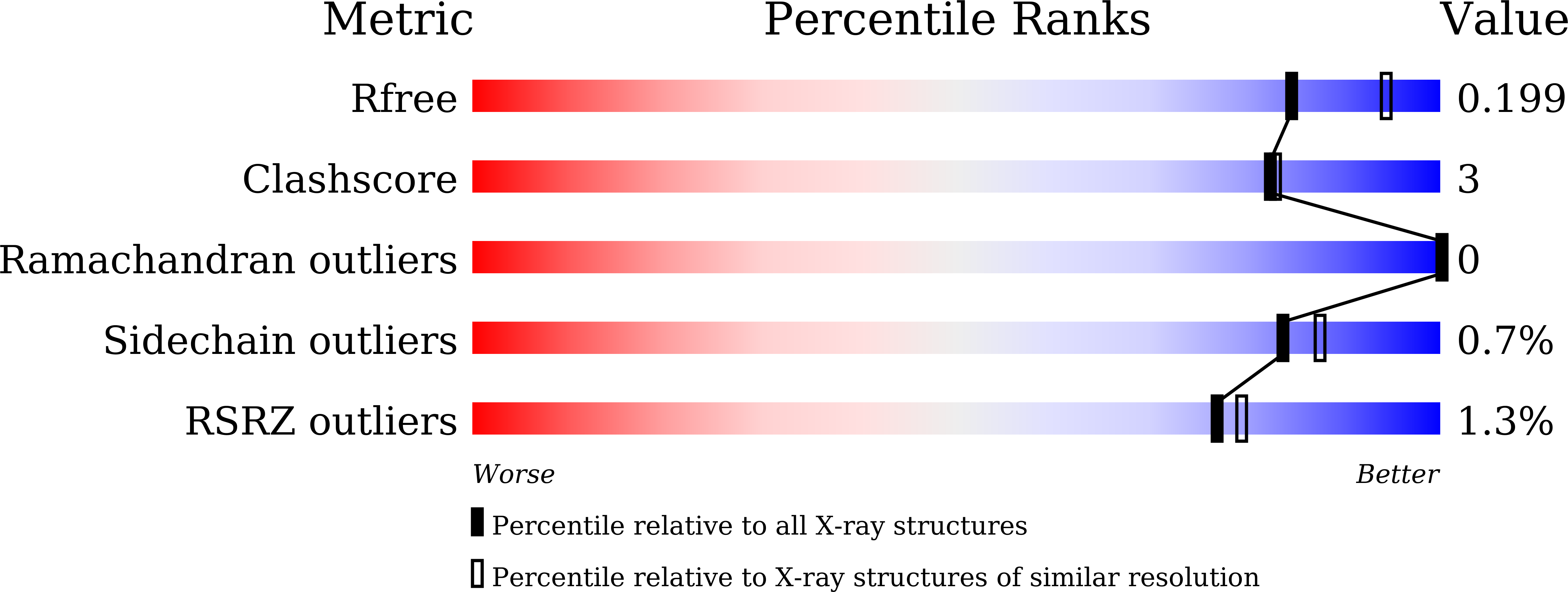
R-Value Free:
0.19
R-Value Work:
0.15
R-Value Observed:
0.15
Space Group:
P 1 21 1