
Deposition Date
2023-10-24
Release Date
2024-09-04
Last Version Date
2024-11-13
Entry Detail
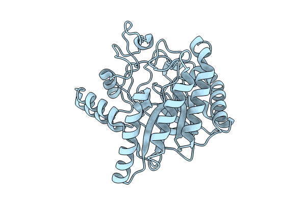
PDB ID:
8WW5
Keywords:
Title:
X-Ray crystal structure of glycoside hydrolase family 6 cellobiohydrolase from Phanerochaete chrysosporium PcCel6A C240S
Biological Source:
Source Organism(s):
Phanerodontia chrysosporium (Taxon ID: 2822231)
Expression System(s):
Method Details:
Experimental Method:
Resolution:
1.01 Å
R-Value Free:
0.15
R-Value Work:
0.14
R-Value Observed:
0.14
Space Group:
P 21 21 21
