8WT3 image
Entry Detail
PDB ID:
8WT3
Keywords:
Title:
Crystal structure of peptidoglycan DL-endopeptidase CwlO
Biological Source:
PDB Version:
Deposition Date:
2023-10-17
Release Date:
2024-07-03
Method Details:
Experimental Method:
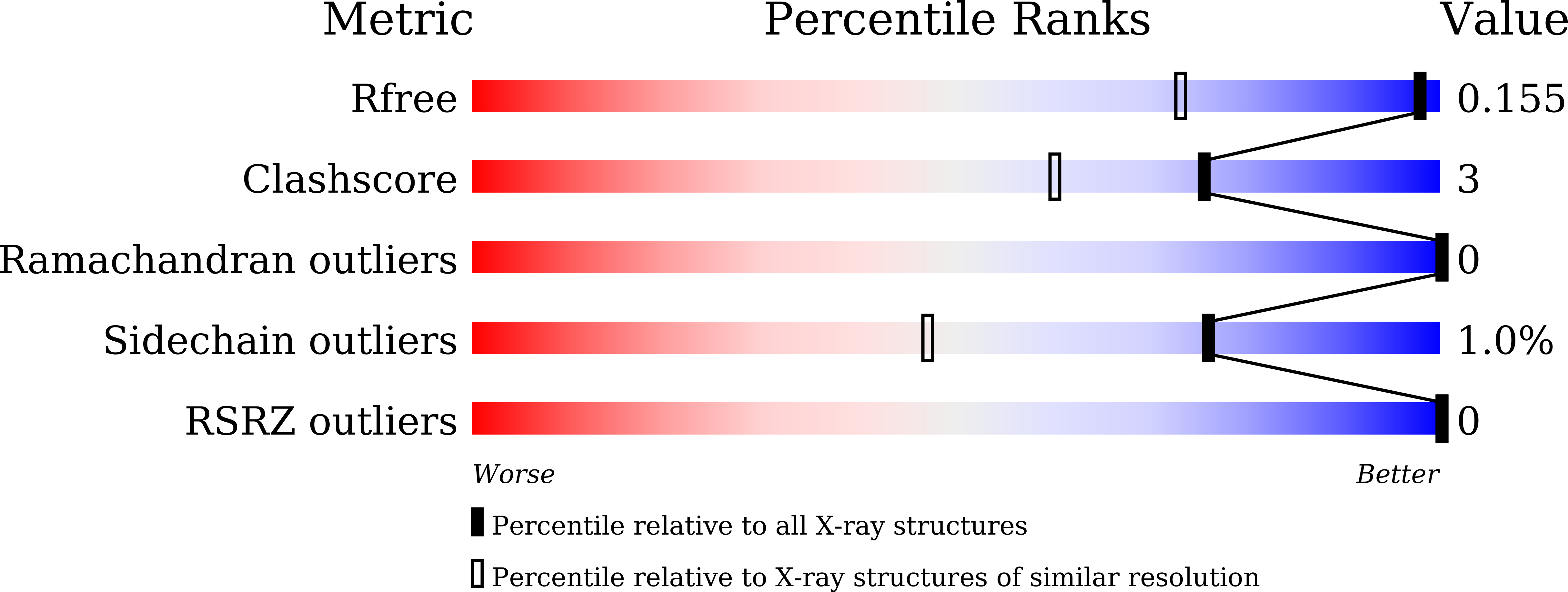
Resolution:
1.20 Å
R-Value Free:
0.15
R-Value Work:
0.14
R-Value Observed:
0.14
Space Group:
P 21 21 21
Macromolecular Entities
Polymer Type:polypeptide(L)
Description:Peptidoglycan DL-endopeptidase CwlO
Chain IDs:A, B
Chain Length:135
Number of Molecules:2
Biological Source:Bacillus subtilis subsp. subtilis str. 168
Ligand Molecules
Primary Citation
Structural analysis of the peptidoglycan DL-endopeptidase CwlO complexed with its inhibitory protein IseA.
Febs J. 291 3723 3736 (2024)
PMID: 38840475 DOI: 10.1111/febs.17197

Abstact

Peptidoglycan DL-endopeptidases locally cleave the peptide stem of peptidoglycan in the bacterial cell wall. This process facilitates bacterial growth and division by loosening the rigid peptidoglycan layer. IseA binds to the active site of multiple DL-endopeptidases and inhibits excessive peptidoglycan degradation that leads to cell lysis. To better understand how IseA inhibits DL-endopeptidase activity, we determined the crystal structure of the peptidoglycan DL-endopeptidase CwlO/IseA complex and compared it with that of the peptidoglycan DL-endopeptidase LytE/IseA complex. Structural analyses showed significant differences between the hydrophobic pocket-binding residues of the DL-endopeptidases (F361 of CwlO and W237 of LytE). Additionally, binding assays showed that the F361 mutation of CwlO to the bulkier hydrophobic residue, tryptophan, increased its binding affinity for IseA, whereas mutation to alanine reduced the affinity. These analyses revealed that the hydrophobic pocket-binding residue of DL-endopeptidases determines IseA-binding affinity and is required for substrate-mimetic inhibition by IseA.

Legend

Protein

Chemical

Disease

Primary Citation of related structures