8WO8 image
Deposition Date 2023-10-06
Release Date 2024-02-14
Last Version Date 2024-06-19
Entry Detail
PDB ID:
8WO8
Title:
Crystal Structure of an RNA-binding protein, FAU-1, from Pyrococcus furiosus
Biological Source:
Source Organism:
Method Details:
Experimental Method:
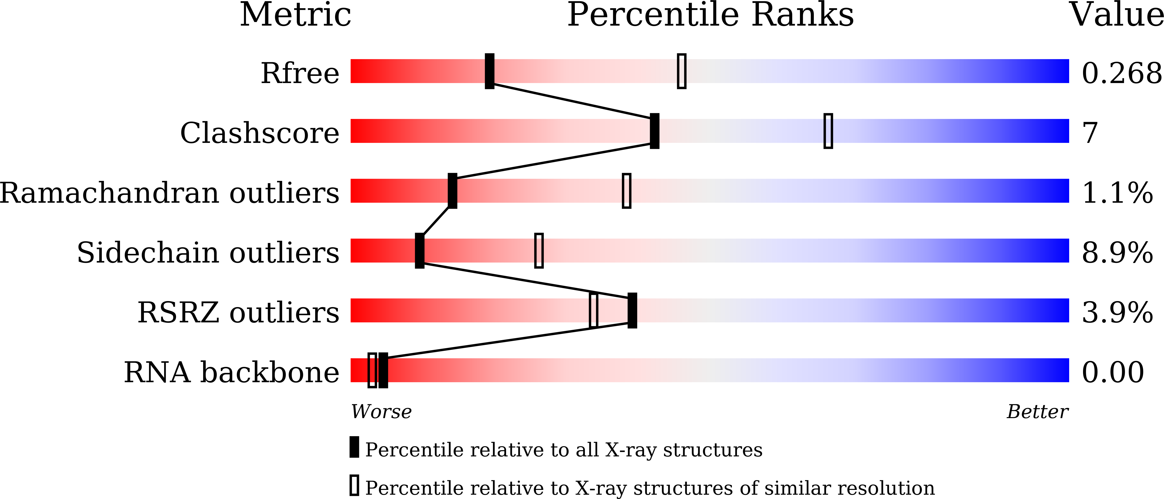
Resolution:
2.78 Å
R-Value Free:
0.26
R-Value Work:
0.20
R-Value Observed:
0.20
Space Group:
P 3 2 1
Macromolecular Entities
Polymer Type:polypeptide(L)
Molecule:Probable ribonuclease FAU-1
Chain IDs:A
Chain Length:479
Number of Molecules:1
Biological Source:Pyrococcus furiosus
Polymer Type:polyribonucleotide
Molecule:RNA (5'-R(P*AP*UP*A)-3')
Chain IDs:B
Chain Length:3
Number of Molecules:1
Biological Source:Escherichia coli
Primary Citation
Homo-trimeric structure of the ribonuclease for rRNA processing, FAU-1, from Pyrococcus furiosus.
J.Biochem. 175 671 676 (2024)
PMID: 38302756 DOI: 10.1093/jb/mvae010

Abstact

Crystal structure of a ribonuclease for ribosomal RNA processing, FAU-1, from Pyrococcus furiosus was determined with the resolution of 2.57 Å in a homo-trimeric form. The monomer structure consists of two domains: N-terminal and C-terminal domains. C-terminal domain forms trimer and each N-terminal domain locates outside of the trimer core. In the obtained crystal, a dinucleotide, pApUp, was bound to the N-terminal domain, indicating that N-terminal domain has the RNA-binding ability. The affinities to RNA of FAU-1 and a fragment corresponding to the N-terminal domain, FAU-ΔC, were confirmed by polyacrylamide gel electrophoresis and nuclear magnetic resonance (NMR). Interestingly, well-dispersed NMR signals were observed at 318K, indicating that the FAU-ΔC-F18 complex form an ordered structure at higher temperature. As predicted in our previous works, FAU-1 and ribonuclease (RNase) E show a structural similarity in their RNA-binding regions. However, structural similarity between RNase E and FAU-1 could be found in the limited regions of the N-terminal domain. On the other hand, structural similarity between C-terminal domain and some proteins including a phosphatase was found. Thus, it is possible that the catalytic site is located in C-terminal domain.

Legend

Protein

Chemical

Disease

Primary Citation of related structures