Deposition Date
2023-09-17
Release Date
2023-12-06
Last Version Date
2024-11-20
Entry Detail
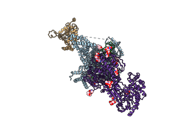
PDB ID:
8WE8
Keywords:
Title:
Human L-type voltage-gated calcium channel Cav1.2 in the presence of calciseptine, amlodipine and pinaverium at 2.9 Angstrom resolution
Biological Source:
Source Organism(s):
Homo sapiens (Taxon ID: 9606)
Dendroaspis polylepis polylepis (Taxon ID: 8620)
Dendroaspis polylepis polylepis (Taxon ID: 8620)
Expression System(s):
Method Details:
Experimental Method:
Resolution:
2.90 Å
Aggregation State:
PARTICLE
Reconstruction Method:
SINGLE PARTICLE