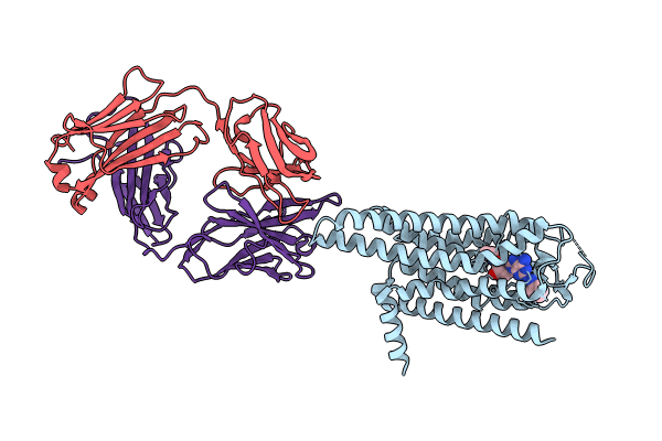
8WDT image
Entry Detail
PDB ID:
8WDT
Title:
Crystal structure of the human adenosine A2A receptor in complex with photoresponsive ligand photoNECA(blue)
Biological Source:
Source Organism:
Host Organism:
PDB Version:
Deposition Date:
2023-09-16
Release Date:
2024-01-17
Method Details:
Experimental Method:
Resolution:
3.34 Å
R-Value Free:
0.26
R-Value Work:
0.21
R-Value Observed:
0.21
Space Group:
C 1 2 1
Macromolecular Entities
Polymer Type:polypeptide(L)
Description:Adenosine receptor A2a
Chain IDs:A
Chain Length:351
Number of Molecules:1
Biological Source:Homo sapiens
Polymer Type:polypeptide(L)
Description:Antibody Fab fragment light chain
Chain IDs:B
Chain Length:213
Number of Molecules:1
Biological Source:Mus musculus
Polymer Type:polypeptide(L)
Description:Antibody Fab fragment heavy chain
Chain IDs:C
Chain Length:224
Number of Molecules:1
Biological Source:Mus musculus
Ligand Molecules
Primary Citation
Crystal structure reveals the binding mode and selectivity of a photoswitchable ligand for the adenosine A 2A receptor.
Biochem.Biophys.Res.Commun. 695 149393 149393 (2023)
PMID: 38171234 DOI: 10.1016/j.bbrc.2023.149393

Abstact

Rational synthetic expansion of photoresponsive ligands is important for photopharmacological studies. Adenosine A2A receptor (A2AR) is stimulated by adenosine and related in Parkinson's disease and other diseases. Here, we report the crystal structure of the A2AR in complex with the novel photoresponsive ligand photoNECA (blue) at 3.34 Å resolution. PhotoNECA (blue) was designed for this structural study and the cell-based assay showed a photoresponsive and receptor selective characteristics of photoNECA (blue) for A2AR. The crystal structure explains the binding mode, photoresponsive mechanism and receptor selectivity of photoNECA (blue). Our study would promote not only the rational design of photoresponsive ligands but also dynamic structural studies of A2AR.

Legend

Protein

Chemical

Disease

Primary Citation of related structures