8WCJ image
Deposition Date 2023-09-12
Release Date 2024-07-31
Last Version Date 2024-07-31
Entry Detail
PDB ID:
8WCJ
Keywords:
Title:
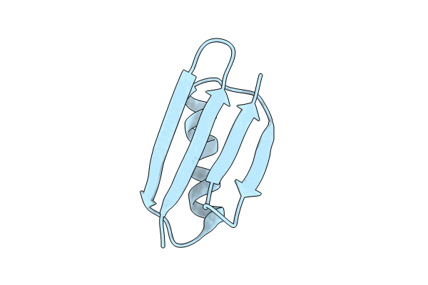
Crystal structure of GB3 penta mutation L5V/K10H/T16S/K19E/Y33I
Biological Source:
Source Organism:
Host Organism:
Method Details:
Experimental Method:
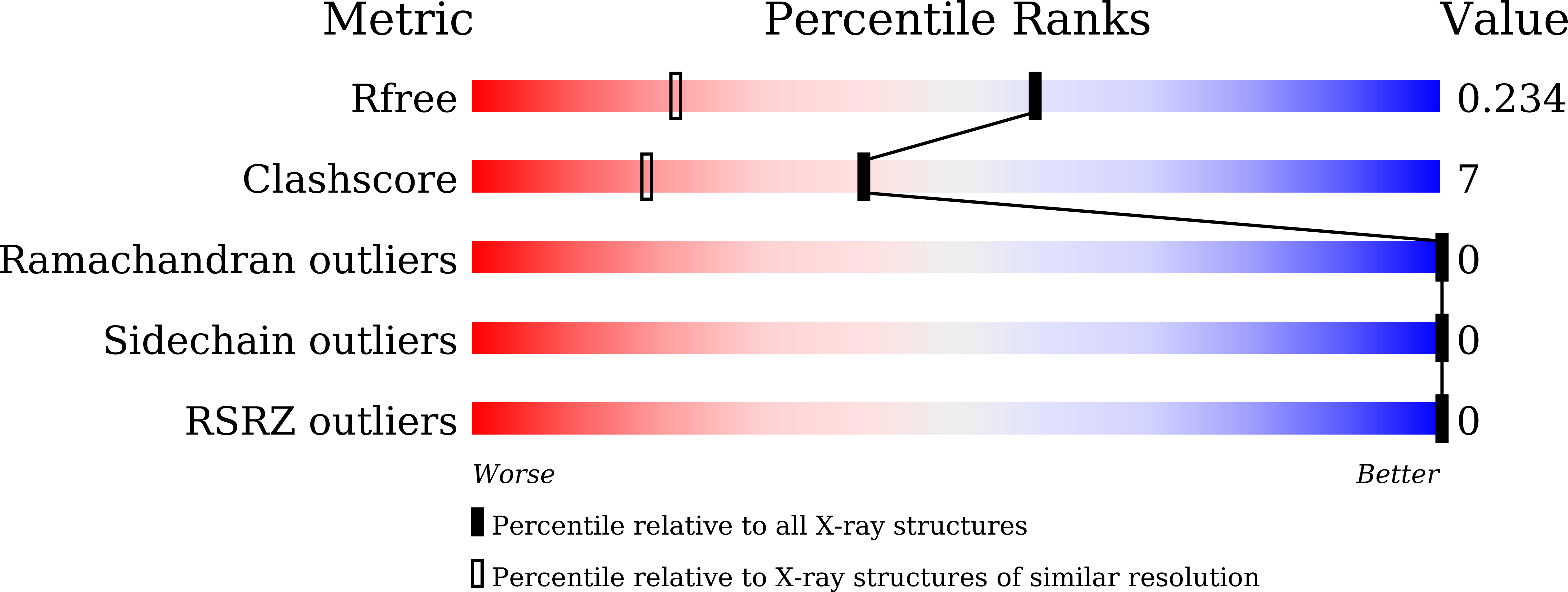
Resolution:
1.55 Å
R-Value Free:
0.23
R-Value Work:
0.20
R-Value Observed:
0.20
Space Group:
I 41 2 2
Macromolecular Entities
Structures with similar UniProt ID
Protein Blast
Polymer Type:polypeptide(L)
Molecule:Immunoglobulin G-binding protein G
Gene (Uniprot):spg
Mutagens:L446V, K451H, T457S, K460E, Y474I
Chain IDs:A
Chain Length:56
Number of Molecules:1
Biological Source:Streptococcus sp. group G
Primary Citation
Protein Allostery Study in Cells Using NMR Spectroscopy.
Anal.Chem. 96 7065 7072 (2024)
PMID: 38652079 DOI: 10.1021/acs.analchem.4c00360

Abstact

Protein allostery is commonly observed in vitro. But how protein allostery behaves in cells is unknown. In this work, a protein monomer-dimer equilibrium system was built with the allosteric effect on the binding characterized using NMR spectroscopy through mutations away from the dimer interface. A chemical shift linear fitting method was developed that enabled us to accurately determine the dissociation constant. A total of 28 allosteric mutations were prepared and grouped to negative allosteric, nonallosteric, and positive allosteric modulators. ∼ 50% of mutations displayed the allosteric-state changes when moving from a buffered solution into cells. For example, there were no positive allosteric modulators in the buffered solution but eight in cells. The change in protein allostery is correlated with the interactions between the protein and the cellular environment. These interactions presumably drive the surrounding macromolecules in cells to transiently bind to the monomer and dimer mutational sites and change the free energies of the two species differently which generate new allosteric effects. These surrounding macromolecules create a new protein allostery pathway that is only present in cells.

Legend

Protein

Chemical

Disease

Primary Citation of related structures