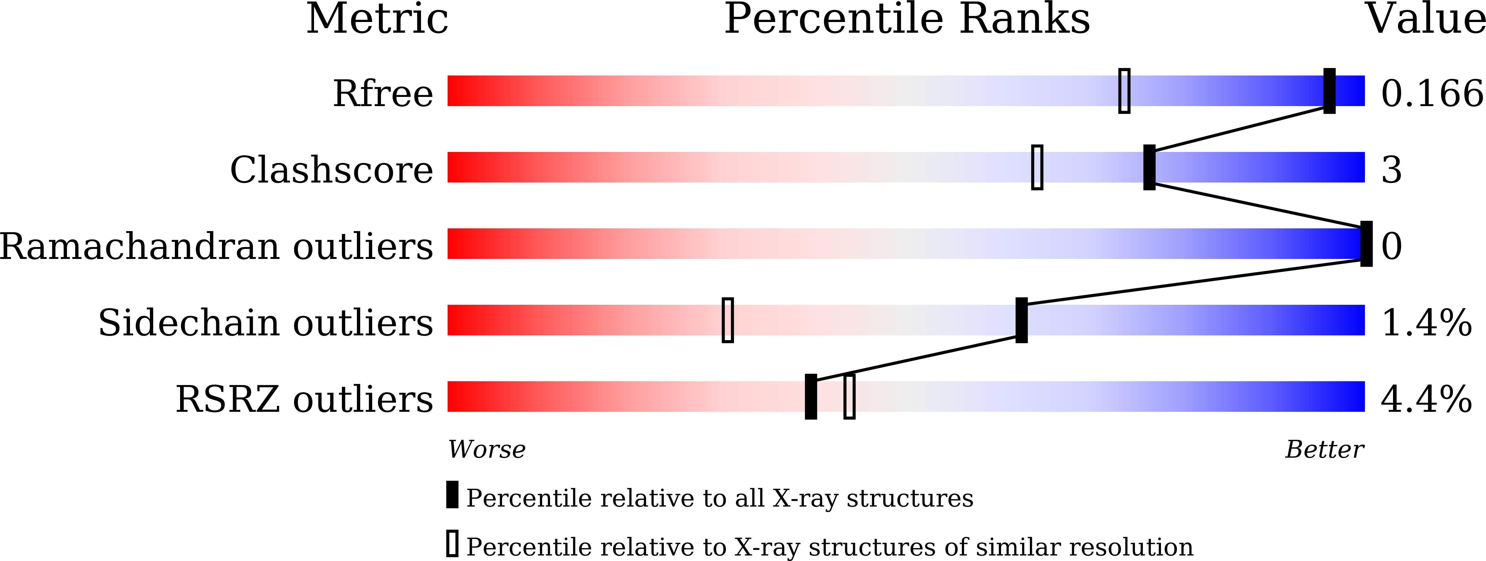
8WCF image
Deposition Date 2023-09-12
Release Date 2024-09-18
Last Version Date 2025-07-02
Entry Detail
PDB ID:
8WCF
Keywords:
Title:
Crystal structure of EcThsB
Biological Source:
Source Organism:
Escherichia coli (Taxon ID: 562)
Host Organism:
Method Details:
Experimental Method:
Resolution:
1.30 Å
R-Value Free:
0.16
R-Value Work:
0.13
R-Value Observed:
0.13
Space Group:
P 21 21 21
Macromolecular Entities
Polymer Type:polypeptide(L)
Molecule:Molecular chaperone Tir
Chain IDs:A
Chain Length:160
Number of Molecules:1
Biological Source:Escherichia coli
Primary Citation
Self-association activates ThsB NAD + hydrolase for defense against phage infection.
Biochem.Biophys.Res.Commun. 776 152217 152217 (2025)
PMID: 40540948 DOI: 10.1016/j.bbrc.2025.152217

Abstact

Toll/interleukin-1 receptor (TIR) domain serves as a canonical component in both animal and plant innate immunity pathways and is indicated, in some cases, to mediate nicotinamide adenine dinucleotide (NAD+) cleavage via self-association. Recent studies have revealed the involvement of TIR domains in a bacterial anti-phage defense system called Thoeris. The Thoeris system consists of two core proteins, ThsA and ThsB. Phage infection triggers the TIR-containing ThsB to produce an isomer of cyclic ADP-ribose, which is then transferred to and activates ThsA, leading to NAD+ depletion and subsequent cell death. However, the mechanism of ThsB activation remains elusive. Here, we present high-resolution crystal structures of E. coli ThsA and ThsB. Notably, an intact NAD+ molecule is observed in the active site of ThsB, implying that monomeric ThsB does not possess NADase activity. We demonstrate that ThsB forms 7-fold oligomers through negative staining electron microscopy, suggesting that self-association activates ThsB NAD+ hydrolase. Our findings indicate a new TIR self-association assembly in bacterial anti-phage systems.

Legend

Protein

Chemical

Disease

Primary Citation of related structures