
Deposition Date
2023-09-06
Release Date
2024-03-06
Last Version Date
2025-07-23
Entry Detail
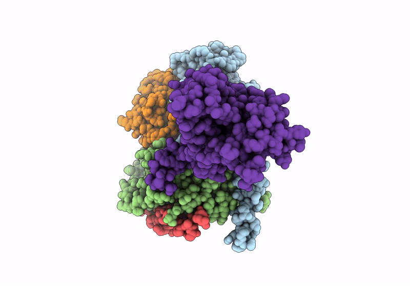
PDB ID:
8WA3
Keywords:
Title:
Cryo-EM structure of peptide free and Gs-coupled GIPR
Biological Source:
Source Organism(s):
Homo sapiens (Taxon ID: 9606)
Bos taurus (Taxon ID: 9913)
Rattus norvegicus (Taxon ID: 10116)
Leptolinea tardivitalis (Taxon ID: 229920)
synthetic construct (Taxon ID: 32630)
Bos taurus (Taxon ID: 9913)
Rattus norvegicus (Taxon ID: 10116)
Leptolinea tardivitalis (Taxon ID: 229920)
synthetic construct (Taxon ID: 32630)
Expression System(s):
Method Details:
Experimental Method:
Resolution:
2.86 Å
Aggregation State:
PARTICLE
Reconstruction Method:
SINGLE PARTICLE
