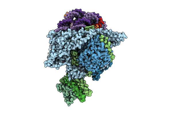
8W9F image
Entry Detail
PDB ID:
8W9F
EMDB ID:
Title:
Cryo-EM structure of the Rpd3S-nucleosome complex from budding yeast in State 3
Biological Source:
PDB Version:
Deposition Date:
2023-09-05
Release Date:
2024-05-15
Method Details:
Experimental Method:
Resolution:
4.40 Å
Aggregation State:
PARTICLE
Reconstruction Method:
SINGLE PARTICLE
Macromolecular Entities
Polymer Type:polypeptide(L)
Description:Transcriptional regulatory protein SIN3
Chain IDs:A
Chain Length:1536
Number of Molecules:1
Biological Source:Saccharomyces cerevisiae
Polymer Type:polypeptide(L)
Description:Histone deacetylase RPD3
Chain IDs:C (auth: B)
Chain Length:433
Number of Molecules:1
Biological Source:Saccharomyces cerevisiae
Polymer Type:polypeptide(L)
Description:Chromatin modification-related protein EAF3
Chain IDs:D (auth: C), E (auth: D), Q (auth: G)
Chain Length:401
Number of Molecules:3
Biological Source:Saccharomyces cerevisiae
Polymer Type:polypeptide(L)
Description:Transcriptional regulatory protein RCO1
Chain IDs:B (auth: E), F
Chain Length:684
Number of Molecules:2
Biological Source:Saccharomyces cerevisiae
Polymer Type:polypeptide(L)
Description:Histone H3.1
Chain IDs:G (auth: a), K (auth: e)
Chain Length:1536
Number of Molecules:2
Biological Source:Homo sapiens
Polymer Type:polypeptide(L)
Description:Histone H4
Chain IDs:H (auth: b), L (auth: f)
Chain Length:433
Number of Molecules:2
Biological Source:Homo sapiens
Polymer Type:polypeptide(L)
Description:Histone H2A type 1-B/E
Chain IDs:I (auth: c), M (auth: g)
Chain Length:401
Number of Molecules:2
Biological Source:Homo sapiens
Polymer Type:polypeptide(L)
Description:Histone H2B type 1-K
Chain IDs:J (auth: d), N (auth: h)
Chain Length:126
Number of Molecules:2
Biological Source:Homo sapiens
Polymer Type:polydeoxyribonucleotide
Description:5-DNA
Chain IDs:O (auth: i)
Chain Length:147
Number of Molecules:1
Biological Source:Homo sapiens
Polymer Type:polydeoxyribonucleotide
Description:3-DNA
Chain IDs:P (auth: j)
Chain Length:147
Number of Molecules:1
Biological Source:Homo sapiens
Ligand Molecules
Primary Citation
Structures and dynamics of Rpd3S complex bound to nucleosome.
Sci Adv 10 eadk7678 eadk7678 (2024)
PMID: 38598631 DOI: 10.1126/sciadv.adk7678

Abstact

The Rpd3S complex plays a pivotal role in facilitating local histone deacetylation in the transcribed regions to suppress intragenic transcription initiation. Here, we present the cryo-electron microscopy structures of the budding yeast Rpd3S complex in both its apo and three nucleosome-bound states at atomic resolutions, revealing the exquisite architecture of Rpd3S to well accommodate a mononucleosome without linker DNA. The Rpd3S core, containing a Sin3 Lobe and two NB modules, is a rigid complex and provides three positive-charged anchors (Sin3_HCR and two Rco1_NIDs) to connect nucleosomal DNA. In three nucleosome-bound states, the Rpd3S core exhibits three distinct orientations relative to the nucleosome, assisting the sector-shaped deacetylase Rpd3 to locate above the SHL5-6, SHL0-1, or SHL2-3, respectively. Our work provides a structural framework that reveals a dynamic working model for the Rpd3S complex to engage diverse deacetylation sites.

Legend

Protein

Chemical

Disease

Primary Citation of related structures