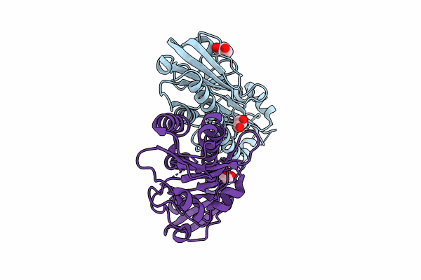
8W7P image
Entry Detail
PDB ID:
8W7P
Title:
Extracellular domain of a sensor histidine kinase
Biological Source:
Source Organism:
Host Organism:
PDB Version:
Deposition Date:
2023-08-31
Release Date:
2024-01-10
Method Details:
Experimental Method:
Resolution:
1.80 Å
R-Value Free:
0.19
R-Value Work:
0.15
R-Value Observed:
0.16
Space Group:
P 1
Macromolecular Entities
Polymer Type:polypeptide(L)
Description:Extracellular domain of a sensor histidine kinase NagS
Chain IDs:A, B
Chain Length:274
Number of Molecules:2
Biological Source:Paenibacillus sp. FPU-7
Ligand Molecules
Primary Citation
Characterization of the extracellular domain of sensor histidine kinase NagS from Paenibacillus sp. str. FPU-7: nagS interacts with oligosaccharide binding protein NagB1 in complexes with N, N'-diacetylchitobiose.
Biosci.Biotechnol.Biochem. 88 294 304 (2024)
PMID: 38059852 DOI: 10.1093/bbb/zbad173

Abstact

We have previously isolated the Gram-positive chitin-degrading bacterium Paenibacillus sp. str. FPU-7. This bacterium traps chitin disaccharide (GlcNAc)2 on its cell surface using two homologous solute-binding proteins, NagB1 and NagB2. Bacteria use histidine kinase (HK) of the two-component regulatory system as an extracellular environment sensor. In this study, we found that nagS, which encodes a HK, is located next to the nagB1 gene. Biochemical experiments revealed that the NagS sensor domain (NagS30-294) interacts with the NagB1-(GlcNAc)2 complex. However, proof of NagS30-294 interacting with NagB1 without (GlcNAc)2 is currently unavailable. In contrast to NagB1, no complex formation was observed between NagS30-294 and NagB2, even in the presence of (GlcNAc)2. The NagS30-294 crystal structure at 1.8 Å resolution suggested that the canonical tandem-Per-Arnt-Sim fold recognizes the NagB1-(GlcNAc)2 complex. This study provides insight into the recognition of chitin oligosaccharides by bacteria.

Legend

Protein

Chemical

Disease

Primary Citation of related structures