8VZ1 image
Entry Detail
PDB ID:
8VZ1
Keywords:
Title:
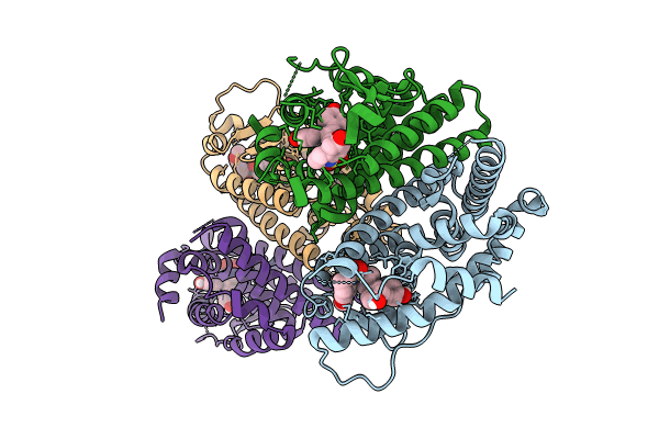
Crystal Structure of the ER-alpha Ligand-binding Domain (L372S, L536S) in complex with k-409
Biological Source:
Source Organism:
PDB Version:
Deposition Date:
2024-02-09
Release Date:
2024-06-12
Method Details:
Experimental Method:
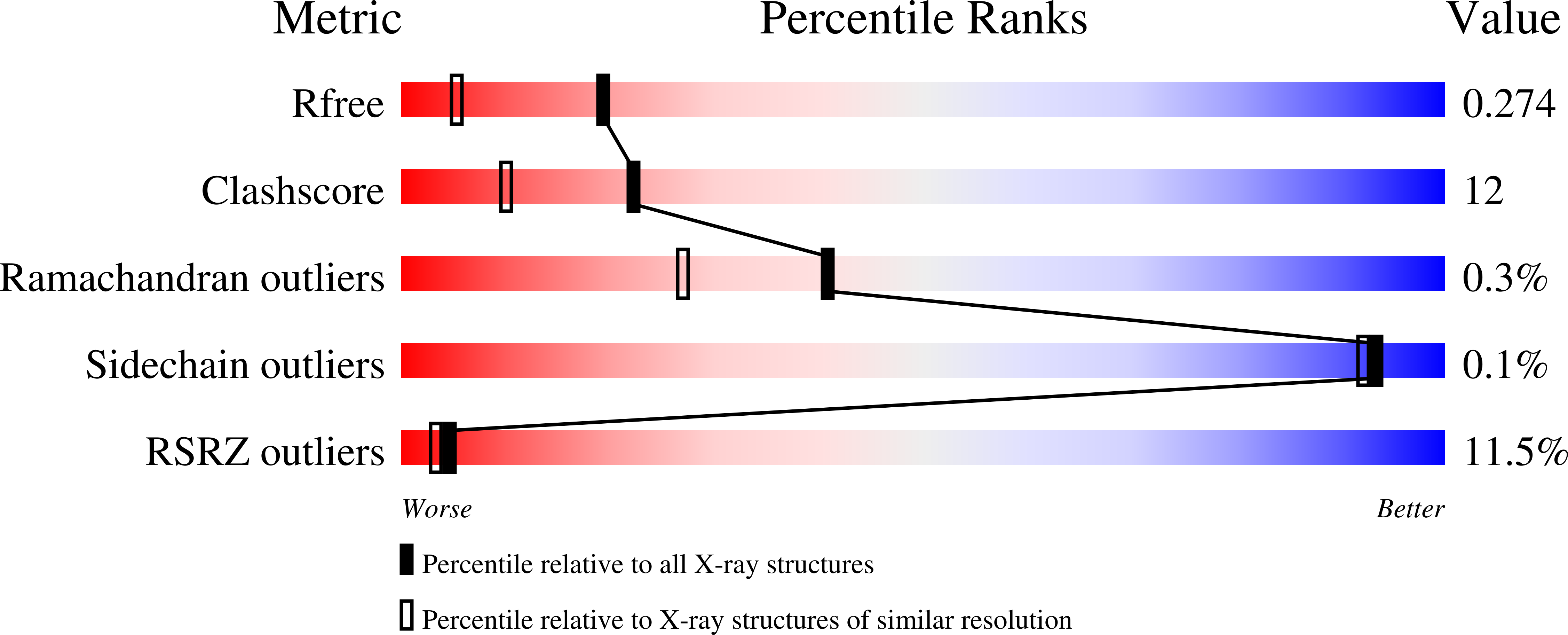
Resolution:
1.82 Å
R-Value Free:
0.27
R-Value Work:
0.22
R-Value Observed:
0.22
Space Group:
P 1
Macromolecular Entities
Polymer Type:polypeptide(L)
Description:Estrogen receptor
Mutations:L372S, L536S
Chain IDs:A, B, C, D
Chain Length:243
Number of Molecules:4
Biological Source:Homo sapiens
Ligand Molecules
Primary Citation
Asymmetric allostery in estrogen receptor-alpha homodimers drives responses to the ensemble of estrogens in the hormonal milieu.
Proc.Natl.Acad.Sci.USA 121 e2321344121 e2321344121 (2024)
PMID: 38830107 DOI: 10.1073/pnas.2321344121

Abstact

The estrogen receptor-α (ER) is thought to function only as a homodimer but responds to a variety of environmental, metazoan, and therapeutic estrogens at subsaturating doses, supporting binding mixtures of ligands as well as dimers that are only partially occupied. Here, we present a series of flexible ER ligands that bind to receptor dimers with individual ligand poses favoring distinct receptor conformations-receptor conformational heterodimers-mimicking the binding of two different ligands. Molecular dynamics simulations showed that the pairs of different ligand poses changed the correlated motion across the dimer interface to generate asymmetric communication between the dimer interface, the ligands, and the surface binding sites for epigenetic regulatory proteins. By examining the binding of the same ligand in crystal structures of ER in the agonist vs. antagonist conformers, we also showed that these allosteric signals are bidirectional. The receptor conformer can drive different ligand binding modes to support agonist vs. antagonist activity profiles, a revision of ligand binding theory that has focused on unidirectional signaling from the ligand to the coregulator binding site. We also observed differences in the allosteric signals between ligand and coregulator binding sites in the monomeric vs. dimeric receptor, and when bound by two different ligands, states that are physiologically relevant. Thus, ER conformational heterodimers integrate two different ligand-regulated activity profiles, representing different modes for ligand-dependent regulation of ER activity.

Legend

Protein

Chemical

Disease

Primary Citation of related structures