8VYL image
Entry Detail
PDB ID:
8VYL
Title:
The structure of Human Hemoglobin in Complex with Nanobody BtNbE11
Biological Source:
Source Organism:
Host Organism:
PDB Version:
Deposition Date:
2024-02-08
Release Date:
2024-07-17
Method Details:
Experimental Method:
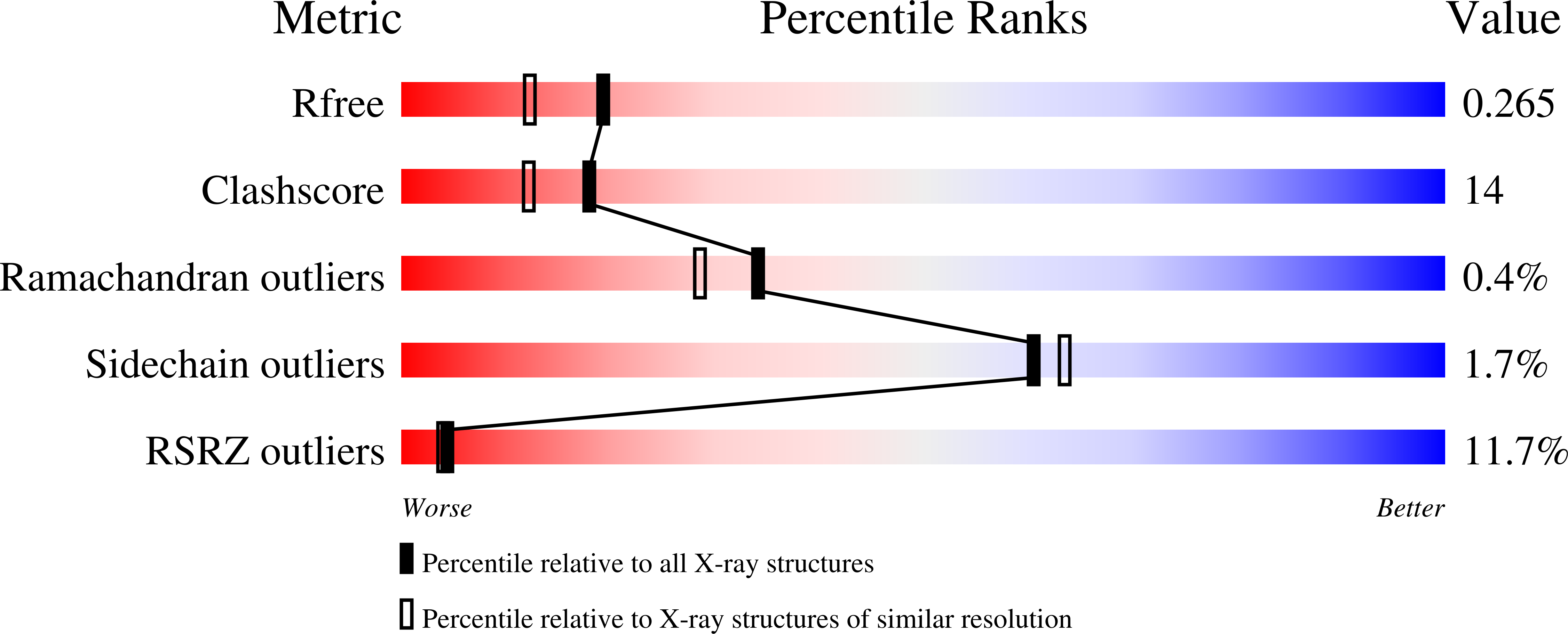
Resolution:
2.02 Å
R-Value Free:
0.26
R-Value Work:
0.20
R-Value Observed:
0.21
Space Group:
C 1 2 1
Macromolecular Entities
Polymer Type:polypeptide(L)
Description:Hemoglobin subunit alpha
Chain IDs:A, B (auth: C)
Chain Length:142
Number of Molecules:2
Biological Source:Homo sapiens
Polymer Type:polypeptide(L)
Description:Hemoglobin subunit beta
Chain IDs:C (auth: B), D
Chain Length:147
Number of Molecules:2
Biological Source:Homo sapiens
Polymer Type:polypeptide(L)
Description:Nanobody BtNbE11
Chain IDs:E (auth: F), F (auth: E)
Chain Length:140
Number of Molecules:2
Biological Source:Escherichia coli
Primary Citation
The structure of a haemoglobin-nanobody complex reveals human beta-subunit-specific interactions.
Febs Lett. 598 2240 2248 (2024)
PMID: 38880764 DOI: 10.1002/1873-3468.14958

Abstact

Haemoglobin (Hb) is a vital oxygen carrier in vertebrates. Low blood Hb levels may indicate anaemia or genetic disorders, while its presence in the lower digestive system suggests colon cancer. Detecting and quantifying human Hb is essential for medical diagnostics. A nanobody-based sandwich-ELISA test was recently developed utilising llama-derived nanobodies NbE11 and NbB9. These nanobodies specifically bind to human Hb without cross-reacting with Hb from other vertebrates. Here, we determine the crystal structure of NbE11 in complex with human Hb. NbE11 binds Hb with high affinity, predominantly binding the β-Hb subunit. Structural differences between human Hb and other vertebrates at the NbE11 binding interface likely explain the assay's lack of cross-reactivity, providing insights for developing Hb binding diagnostics.

Legend

Protein

Chemical

Disease

Primary Citation of related structures