8VY2 image
Deposition Date 2024-02-06
Release Date 2025-05-07
Last Version Date 2025-08-20
Entry Detail
PDB ID:
8VY2
Title:
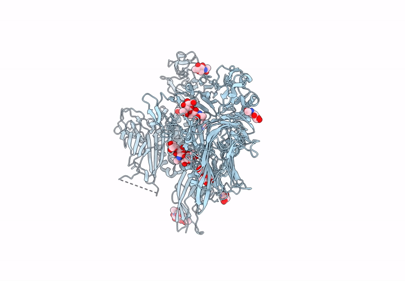
Structure of mCELSR1 extracellular region containing CADH9-GAIN domains
Biological Source:
Source Organism:
Mus musculus (Taxon ID: 10090)
Host Organism:
Method Details:
Experimental Method:
Resolution:
4.30 Å
Aggregation State:
PARTICLE
Reconstruction Method:
SINGLE PARTICLE
Macromolecular Entities
Structures with similar UniProt ID
Protein Blast
Polymer Type:polypeptide(L)
Molecule:Cadherin EGF LAG seven-pass G-type receptor 1
Gene (Uniprot):Celsr1
Chain IDs:A
Chain Length:2232
Number of Molecules:1
Biological Source:Mus musculus
Ligand Molecules
Primary Citation
Structural basis for regulation of CELSR1 by a compact module in its extracellular region.
Nat Commun 16 3972 3972 (2025)
PMID: 40295529 DOI: 10.1038/s41467-025-59319-8

Abstact

The Cadherin EGF Laminin G seven-pass G-type receptor subfamily (CELSR/ADGRC) is one of the most conserved among adhesion G protein-coupled receptors and is essential for animal development. The extracellular regions (ECRs) of CELSRs are large with 23 adhesion domains. However, molecular insight into CELSR function is sparsely available. Here, we report the 3.8 Å cryo-EM reconstruction of the mouse CELSR1 ECR and reveal that 14 domains form a compact module mediated by conserved interactions majorly between the CADH9 and C-terminal GAIN domains. In the presence of Ca2+, the CELSR1 ECR forms a dimer species mediated by the cadherin repeats putatively in an antiparallel fashion. Cell-based assays reveal the N-terminal CADH1-8 repeat is required for cell-cell adhesion and the C-terminal CADH9-GAIN compact module can regulate cellular adhesion. Our work provides molecular insight into how one of the largest GPCRs uses defined structural modules to regulate receptor function.

Legend

Protein

Chemical

Disease

Primary Citation of related structures
Feedback Form
Name
Email
Institute
Feedback