8VXN image
Entry Detail
PDB ID:
8VXN
Keywords:
Title:
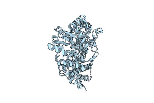
Human Bcl-2/Bcl-xL Chimera Fused to Maltose-Binding Protein
Biological Source:
PDB Version:
Deposition Date:
2024-02-05
Release Date:
2024-10-02
Method Details:
Experimental Method:
Resolution:
2.09 Å
R-Value Free:
0.26
R-Value Work:
0.20
R-Value Observed:
0.20
Space Group:
P 21 21 21
Macromolecular Entities
Polymer Type:polypeptide(L)
Description:Maltose/maltodextrin-binding periplasmic protein fused to apoptosis regulator Bcl-2/Bcl-xL chimera
Chain IDs:A
Chain Length:547
Number of Molecules:1
Biological Source:Escherichia coli (strain K12), Homo sapiens
Primary Citation
Hydrogen/Deuterium Exchange and Protein Oxidative Footprinting with Mass Spectrometry Collectively Discriminate the Binding of Small-Molecule Therapeutics to Bcl-2.
Anal.Chem. 97 4329 4340 (2025)
PMID: 39969248 DOI: 10.1021/acs.analchem.4c04516

Abstact

Characterizing protein-ligand interactions is crucial to understanding cellular metabolism and guiding drug discovery and development. Herein, we explore complementing hydrogen/deuterium exchange mass spectrometry (HDX-MS) with a recently developed Fenton chemistry-based approach to protein oxidative footprinting mass spectrometry (OX-MS) to discriminate the binding of small-molecule therapeutics. Using drug-dependent perturbation as the experimental report, this combination of techniques more clearly differentiates the in-solution binding profiles of Venetoclax (ABT-199, GDC-0199-AbbVie and Genentech) and a drug candidate S55746 (Servier) to the apoptotic regulatory protein Bcl-2 than either technique alone. These results highlight the value of combining these methods to compare compounds in drug discovery and development. To better understand the structural context of the HDX-MS and OX-MS drug-dependent perturbations, we mapped these data on Bcl-2-Venetoclax and Bcl-2-S55746 cocrystal structures and compared these results with the structure of apo Bcl-2. HDX-MS shows that Venetoclax more strongly impacts the protein backbone compared to S55746. OX-MS reveals oxidation perturbations rationalized by direct side-chain protection as well as by crystallographically observed drug-induced protein restructuring. Both methods report the perturbation of some, but not all, residues mapped within 4 Å of the bound drugs in the crystal structures. Concordant characterization of backbone and side-chain accessibility will enhance our understanding of in-solution protein structure dynamics and protein-ligand interactions during drug discovery, development, and characterization, particularly when high-resolution structures are lacking.

Legend

Protein

Chemical

Disease

Primary Citation of related structures