8VRI image
Entry Detail
PDB ID:
8VRI
Keywords:
Title:
E. coli peptidyl-prolyl cis-trans isomerase containing difluoro-leucines
Biological Source:
Source Organism:
Host Organism:
PDB Version:
Deposition Date:
2024-01-22
Release Date:
2024-05-22
Method Details:
Experimental Method:
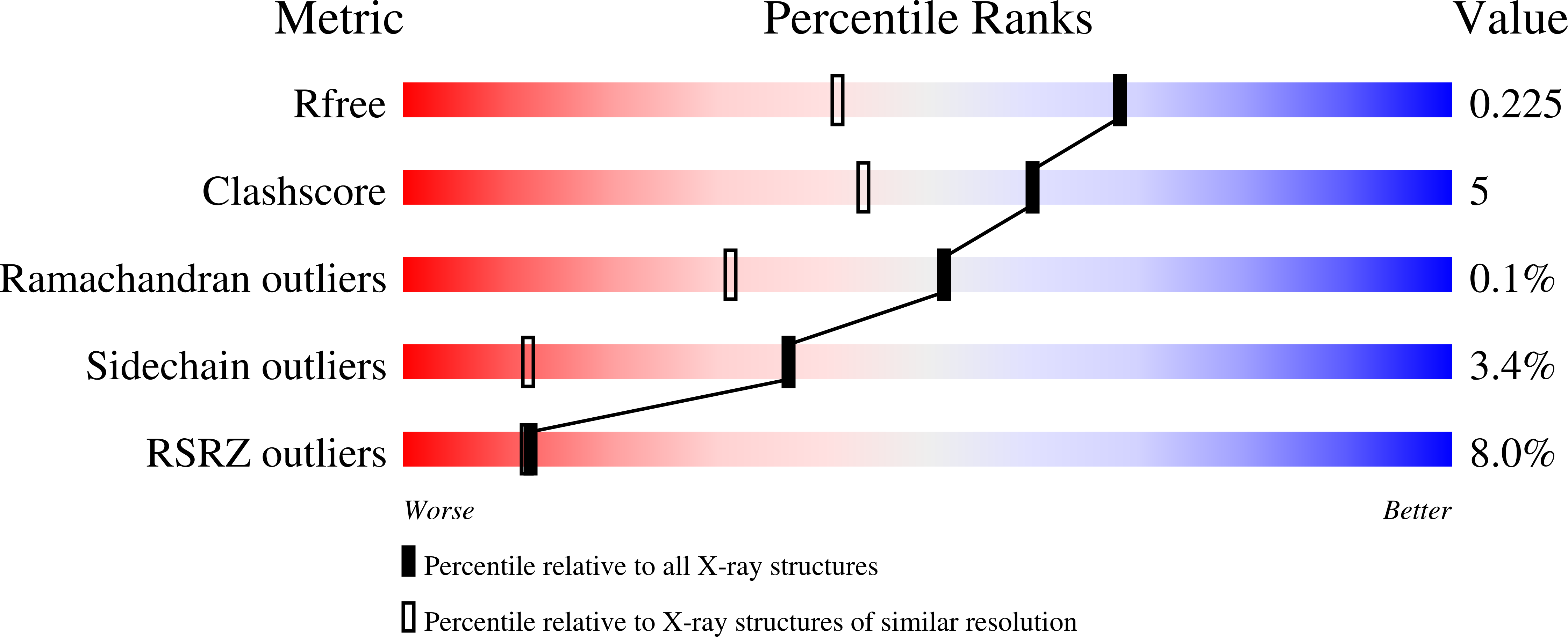
Resolution:
1.65 Å
R-Value Free:
0.22
R-Value Work:
0.18
R-Value Observed:
0.18
Space Group:
P 21 21 21
Macromolecular Entities
Polymer Type:polypeptide(L)
Description:Peptidyl-prolyl cis-trans isomerase B
Chain IDs:A, B
Chain Length:170
Number of Molecules:2
Biological Source:Escherichia coli
Polymer Type:polypeptide(L)
Description:Peptidyl-prolyl cis-trans isomerase B
Chain IDs:C, D, E
Chain Length:170
Number of Molecules:3
Biological Source:Escherichia coli
Modified Residue
Compound ID Chain ID Parent Comp ID Details 2D Image
FFL A LEU modified residue
FME A MET modified residue
Primary Citation
Conformational Preferences of the Non-Canonical Amino Acids (2 S ,4 S )-5-Fluoroleucine, (2 S ,4 R )-5-Fluoroleucine, and 5,5'-Difluoroleucine in a Protein.
Biochemistry 63 1388 1394 (2024)
PMID: 38742763 DOI: 10.1021/acs.biochem.4c00081

Abstact

Proteins produced with leucine analogues, where CH2F groups substitute specific methyl groups, can readily be probed by 19F NMR spectroscopy. As CF and CH groups are similar in hydrophobicity and size, fluorinated leucines are expected to cause minimal structural perturbation, but the impact of fluorine on the rotational freedom of CH2F groups is unclear. We present high-resolution crystal structures of Escherichia coli peptidyl-prolyl cis-trans isomerase B (PpiB) prepared with uniform high-level substitution of leucine by (2S,4S)-5-fluoroleucine, (2S,4R)-5-fluoroleucine, or 5,5'-difluoroleucine. Apart from the fluorinated leucine residues, the structures show complete structural conservation of the protein backbone and the amino acid side chains except for a single isoleucine side chain located next to a fluorine atom in the hydrophobic core of the protein. The carbon skeletons of the fluorinated leucine side chains are also mostly conserved. The CH2F groups show a strong preference for staggered rotamers and often appear locked into single rotamers. Substitution of leucine CH3 groups for CH2F groups is thus readily tolerated in the three-dimensional (3D) structure of a protein, and the rotation of CH2F groups can be halted at cryogenic temperatures.

Legend

Protein

Chemical

Disease

Primary Citation of related structures