8VK4 image
Deposition Date 2024-01-08
Release Date 2024-02-07
Last Version Date 2024-10-09
Entry Detail
PDB ID:
8VK4
Title:
Structure of mouse RyR1 in complex with S100A1 (high-Ca2+/CFF/ATP dataset)
Biological Source:
Source Organism:
Mus musculus (Taxon ID: 10090)
Host Organism:
Method Details:
Experimental Method:
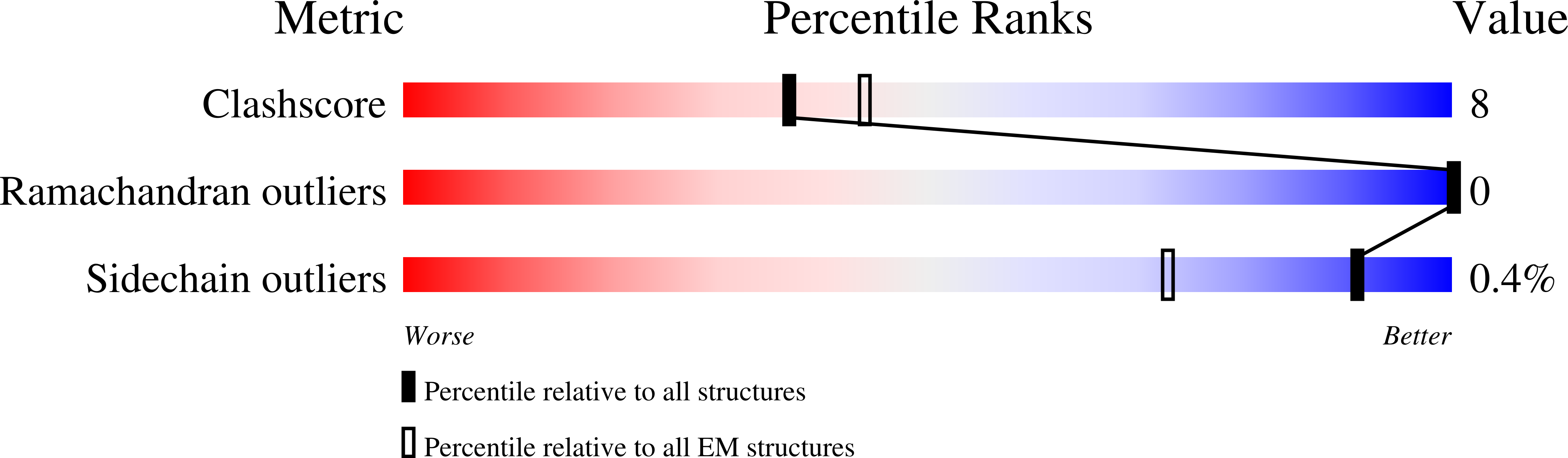
Resolution:
3.56 Å
Aggregation State:
PARTICLE
Reconstruction Method:
SINGLE PARTICLE
Macromolecular Entities
Polymer Type:polypeptide(L)
Molecule:Ryanodine receptor 1
Gene (Uniprot):Ryr1
Chain IDs:G (auth: D), H (auth: A), I (auth: B), J (auth: C)
Chain Length:5035
Number of Molecules:4
Biological Source:Mus musculus
Polymer Type:polypeptide(L)
Molecule:Peptidyl-prolyl cis-trans isomerase FKBP1A
Gene (Uniprot):Fkbp1a
Chain IDs:A (auth: E), D (auth: F), E (auth: G), F (auth: H)
Chain Length:108
Number of Molecules:4
Biological Source:Mus musculus
Polymer Type:polypeptide(L)
Molecule:Protein S100A1
Chain IDs:B (auth: I), C (auth: J), K, L, M (auth: N), N (auth: M), O, P
Chain Length:94
Number of Molecules:8
Biological Source:Mus musculus
Primary Citation
Structural insights into the regulation of RyR1 by S100A1.
Proc.Natl.Acad.Sci.USA 121 e2400497121 e2400497121 (2024)
PMID: 38917010 DOI: 10.1073/pnas.2400497121

Abstact

S100A1, a small homodimeric EF-hand Ca2+-binding protein (~21 kDa), plays an important regulatory role in Ca2+ signaling pathways involved in various biological functions including Ca2+ cycling and contractile performance in skeletal and cardiac myocytes. One key target of the S100A1 interactome is the ryanodine receptor (RyR), a huge homotetrameric Ca2+ release channel (~2.3 MDa) of the sarcoplasmic reticulum. Here, we report cryoelectron microscopy structures of S100A1 bound to RyR1, the skeletal muscle isoform, in absence and presence of Ca2+. Ca2+-free apo-S100A1 binds beneath the bridging solenoid (BSol) and forms contacts with the junctional solenoid and the shell-core linker of RyR1. Upon Ca2+-binding, S100A1 undergoes a conformational change resulting in the exposure of the hydrophobic pocket known to serve as a major interaction site of S100A1. Through interactions of the hydrophobic pocket with RyR1, Ca2+-bound S100A1 intrudes deeper into the RyR1 structure beneath BSol than the apo-form and induces sideways motions of the C-terminal BSol region toward the adjacent RyR1 protomer resulting in tighter interprotomer contacts. Interestingly, the second hydrophobic pocket of the S100A1-dimer is largely exposed at the hydrophilic surface making it prone to interactions with the local environment, suggesting that S100A1 could be involved in forming larger heterocomplexes of RyRs with other protein partners. Since S100A1 interactions stabilizing BSol are implicated in the regulation of RyR-mediated Ca2+ release, the characterization of the S100A1 binding site conserved between RyR isoforms may provide the structural basis for the development of therapeutic strategies regarding treatments of RyR-related disorders.

Legend

Protein

Chemical

Disease

Primary Citation of related structures