8VIZ image
Deposition Date 2024-01-05
Release Date 2024-10-16
Last Version Date 2025-06-04
Entry Detail
PDB ID:
8VIZ
Keywords:
Title:
Structure of full-length gelsolin bound to the barbed end of F-actin
Biological Source:
Source Organism:
Host Organism:
Method Details:
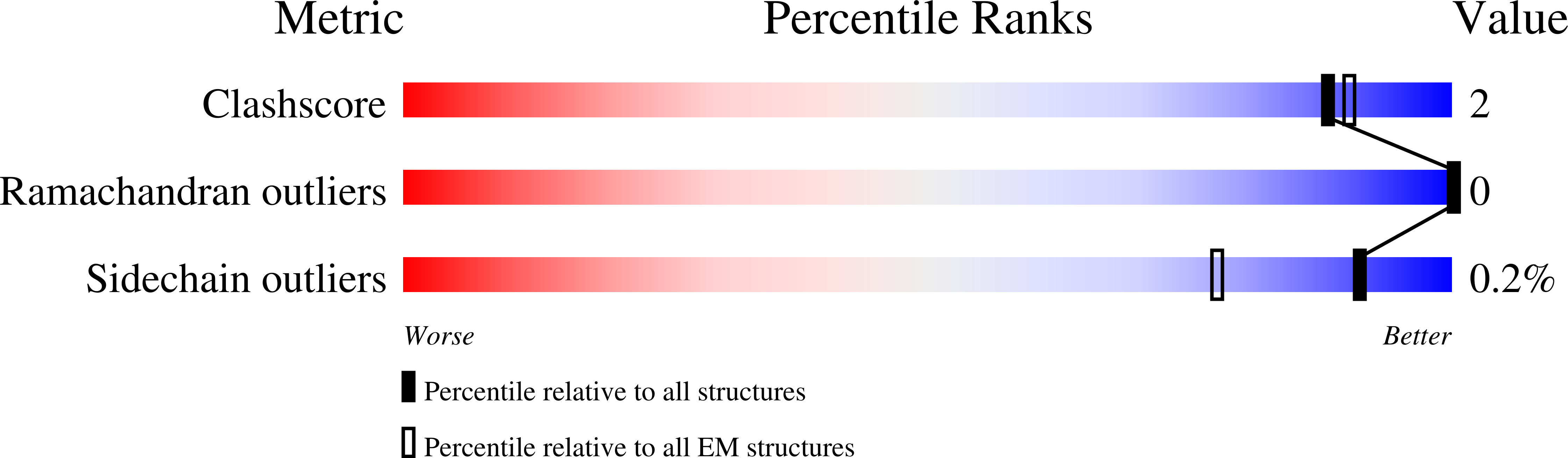
Experimental Method:
Resolution:
2.63 Å
Aggregation State:
PARTICLE
Reconstruction Method:
SINGLE PARTICLE
Macromolecular Entities
Structures with similar UniProt ID
Protein Blast
Polymer Type:polypeptide(L)
Molecule:Actin, alpha skeletal muscle
Gene (Uniprot):ACTA1
Chain IDs:A, B, C, D, E, F
Chain Length:375
Number of Molecules:6
Biological Source:Oryctolagus cuniculus
Structures with similar UniProt ID
Protein Blast
Polymer Type:polypeptide(L)
Molecule:Gelsolin
Gene (Uniprot):GSN
Chain IDs:G
Chain Length:746
Number of Molecules:1
Biological Source:Homo sapiens
Modified Residue
Compound ID Chain ID Parent Comp ID Details 2D Image
HIC A HIS modified residue
Primary Citation
Mechanism of actin filament severing and capping by gelsolin.
Nat.Struct.Mol.Biol. 32 237 242 (2025)
PMID: 39448849 DOI: 10.1038/s41594-024-01412-5

Abstact

Gelsolin is the prototypical member of a family of Ca2+-activated F-actin severing and capping proteins. Here we report structures of Ca2+-bound human gelsolin at the barbed end of F-actin. One structure reveals gelsolin's six domains (G1G6) and interdomain linkers wrapping around F-actin, while another shows domains G1G3-a fragment observed during apoptosis-binding on both sides of F-actin. Conformational changes that trigger severing occur on one side of F-actin with G1G6 and on both sides with G1G3. Gelsolin remains bound after severing, blocking subunit exchange.

Legend

Protein

Chemical

Disease

Primary Citation of related structures
Feedback Form
Name
Email
Institute
Feedback