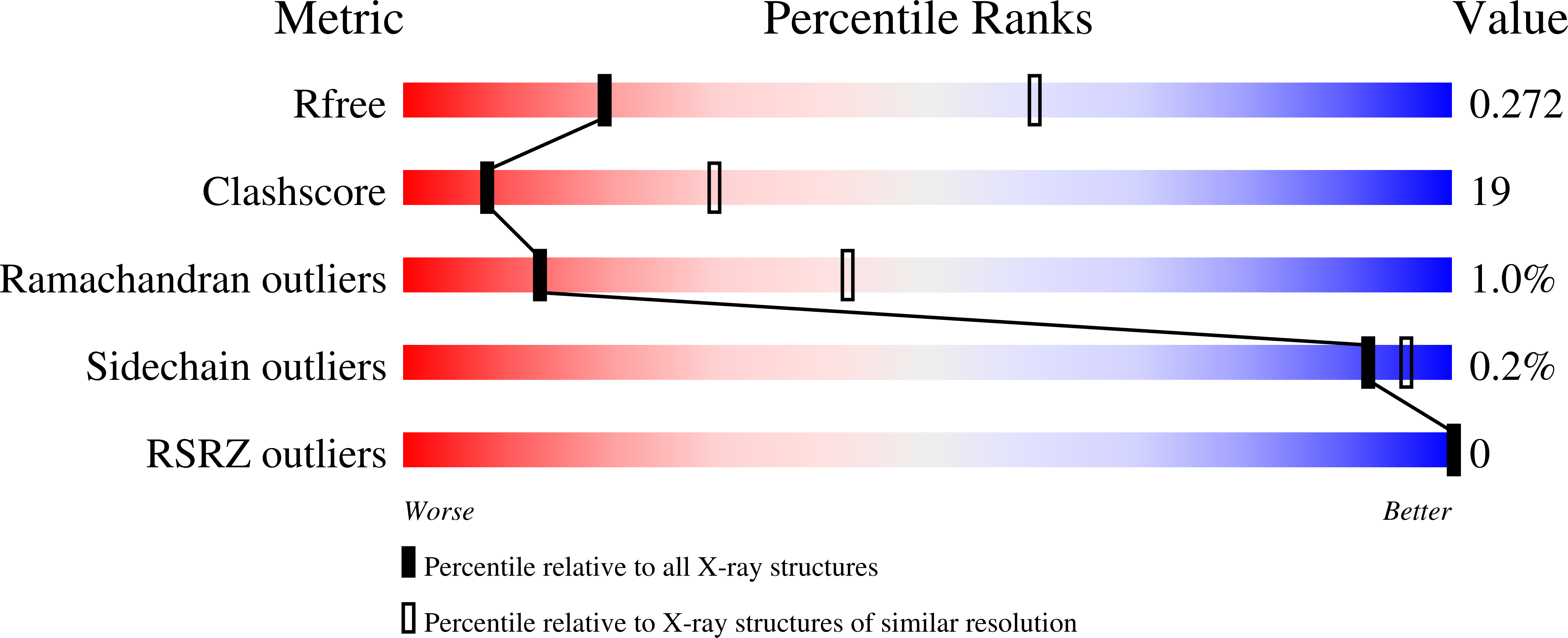
8VBS image
Deposition Date 2023-12-12
Release Date 2024-10-16
Last Version Date 2024-11-27
Entry Detail
PDB ID:
8VBS
Keywords:
Title:
E. coli cysteine desulfurase SufS bound to SufE C51A
Biological Source:
Source Organism:
Escherichia coli (Taxon ID: 562)
Host Organism:
Method Details:
Experimental Method:
Resolution:
3.31 Å
R-Value Free:
0.27
R-Value Work:
0.23
R-Value Observed:
0.23
Space Group:
P 21 21 2
Macromolecular Entities
Structures with similar UniProt ID
Protein Blast
Polymer Type:polypeptide(L)
Molecule:Cysteine desulfurase
Gene (Uniprot):sufS
Chain IDs:A, C (auth: B)
Chain Length:406
Number of Molecules:2
Biological Source:Escherichia coli
Structures with similar UniProt ID
Protein Blast
Polymer Type:polypeptide(L)
Molecule:Cysteine desulfuration protein SufE
Mutagens:C51A, E107C
Chain IDs:B (auth: C), D
Chain Length:138
Number of Molecules:2
Biological Source:Escherichia coli
Modified Residue
Compound ID Chain ID Parent Comp ID Details 2D Image
CSS A CYS modified residue
LLP A LYS modified residue
Ligand Molecules
Primary Citation
The structure of the SufS-SufE complex reveals interactions driving protected persulfide transfer in iron-sulfur cluster biogenesis.
J.Biol.Chem. 300 107641 107641 (2024)
PMID: 39122000 DOI: 10.1016/j.jbc.2024.107641

Abstact

Fe-S clusters are critical cofactors for redox chemistry in all organisms. The cysteine desulfurase, SufS, provides sulfur in the SUF Fe-S cluster bioassembly pathway. SufS is a dimeric, pyridoxal 5'-phosphate-dependent enzyme that uses cysteine as a substrate to generate alanine and a covalent persulfide on an active site cysteine residue. SufS enzymes are activated by an accessory transpersulfurase protein, either SufE or SufU depending on the organism, which accepts the persulfide product and delivers it to downstream partners for Fe-S assembly. Here, using Escherichia coli proteins, we present the first X-ray crystal structure of a SufS/SufE complex. There is a 1:1 stoichiometry with each monomeric unit of the EcSufS dimer bound to one EcSufE subunit, though one EcSufE is rotated ∼7° closer to the EcSufS active site. EcSufE makes clear interactions with the α16 helix of EcSufS and site-directed mutants of several α16 residues were deficient in EcSufE binding. Analysis of the EcSufE structure showed a loss of electron density at the EcSufS/EcSufE interface for a flexible loop containing the highly conserved residue R119. An R119A EcSufE variant binds EcSufS but is not active in cysteine desulfurase assays and fails to support Fe-S cluster bioassembly in vivo. 35S-transfer assays suggest that R119A EcSufE can receive a persulfide, suggesting the residue may function in a release mechanism. The structure of the EcSufS/EcSufE complex allows for comparison with other cysteine desulfurases to understand mechanisms of protected persulfide transfer across protein interfaces.

Legend

Protein

Chemical

Disease

Primary Citation of related structures