8V9P image
Deposition Date 2023-12-08
Release Date 2023-12-20
Last Version Date 2025-02-12
Entry Detail
PDB ID:
8V9P
Keywords:
Title:
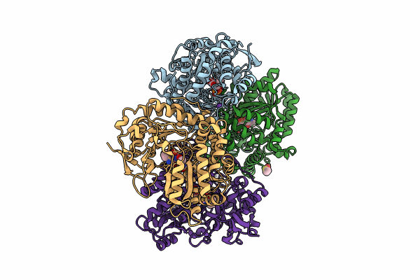
Proteus vulgaris tryptophan indole-lyase complexed with (3S)-dioxindolyl-L-alanine
Biological Source:
Source Organism:
Proteus vulgaris (Taxon ID: 585)
Method Details:
Experimental Method:
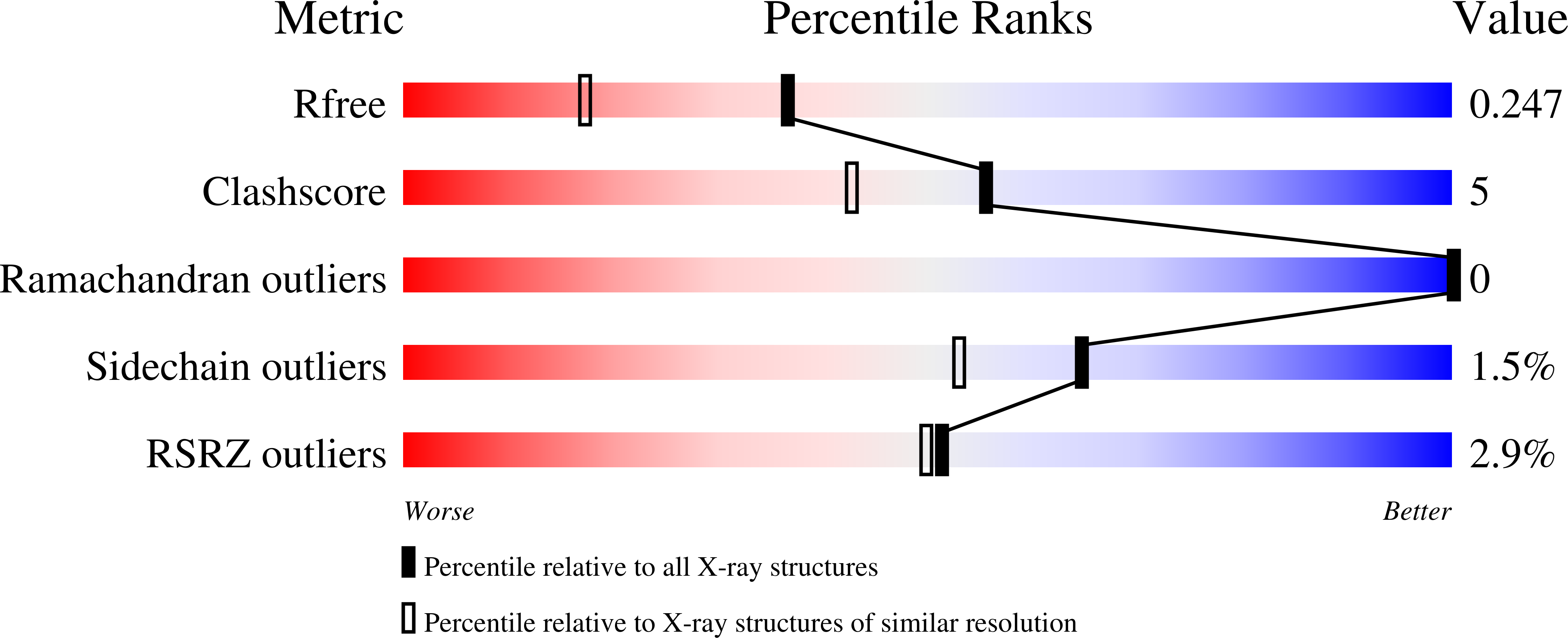
Resolution:
1.85 Å
R-Value Free:
0.24
R-Value Work:
0.19
R-Value Observed:
0.20
Space Group:
P 1 21 1
Macromolecular Entities
Structures with similar UniProt ID
Protein Blast
Polymer Type:polypeptide(L)
Molecule:Tryptophanase
Gene (Uniprot):tnaA
Chain IDs:A, B, C, D
Chain Length:467
Number of Molecules:4
Biological Source:Proteus vulgaris
Primary Citation
Structural Snapshots of Proteus vulgaris Tryptophan Indole-Lyase Reveal Insights into the Catalytic Mechanism.
Acs Catalysis 14 11498 11511 (2024)
PMID: 39114092 DOI: 10.1021/acscatal.4c03232

Abstact

Tryptophan indole lyase (TIL; [E.C. 4.1.99.1]) is a bacterial pyridoxal-5'-phosphate (PLP)-dependent enzyme that catalyzes reversible β-elimination of indole from L-tryptophan. The mechanism of elimination of indole from L-tryptophan starts with the formation of an external aldimine of the substrate and PLP, followed by deprotonation of the α-CH of the substrate, forming a resonance-stabilized quinonoid intermediate. Proton transfer to C3 of the indole ring and carbon-carbon bond cleavage of the quinonoid intermediate provide indole and aminoacrylate bound to PLP, which then releases indole, followed by iminopyruvate. We have now determined the X-ray crystal structures of TIL complexes with (3S)-dioxindolyl-l-alanine, an inhibitor, and with substrates L-tryptophan, 7-aza-L-tryptophan, and S-ethyl-l-cysteine (SEC) in the presence of benzimidazole (BZI), an isostere of the product indole. These structures show a mixture of gem-diamine, external aldimine, quinonoid, and aminoacrylate intermediates, in both open and closed active site conformations. In the closed conformations of L-tryptophan, (3S)-dioxindolyl-l-alanine, and 7-aza-L-tryptophan complexes, hydrogen bonds form between Asp-133 with N1 of the ligand heterocyclic ring and NE2 of His-458 in the small domain of TIL. This hydrogen bond also forms in the BZI complex with the aminoacrylate intermediates formed from both L-tryptophan and SEC. The closed quinonoid complex of 7-aza-L-tryptophan shows that the azaindole ring in the closed conformation is bent out of plane of the Cβ-C3 bond by about 40°, putting it in a geometry that leads toward the transition-state geometry. Thus, both conformational dynamics and substrate activation play critical roles in the reaction mechanism of the TIL.

Legend

Protein

Chemical

Disease

Primary Citation of related structures
Feedback Form
Name
Email
Institute
Feedback