8V5L image
Deposition Date 2023-11-30
Release Date 2024-10-09
Last Version Date 2024-11-13
Entry Detail
PDB ID:
8V5L
Keywords:
Title:
Structure of the Varicella Zoster Virus (VZV) gI binding domain of glycoprotein E (gE) in complex with human Fab 1A2 and 1E12
Biological Source:
Source Organism:
Host Organism:
Method Details:
Experimental Method:
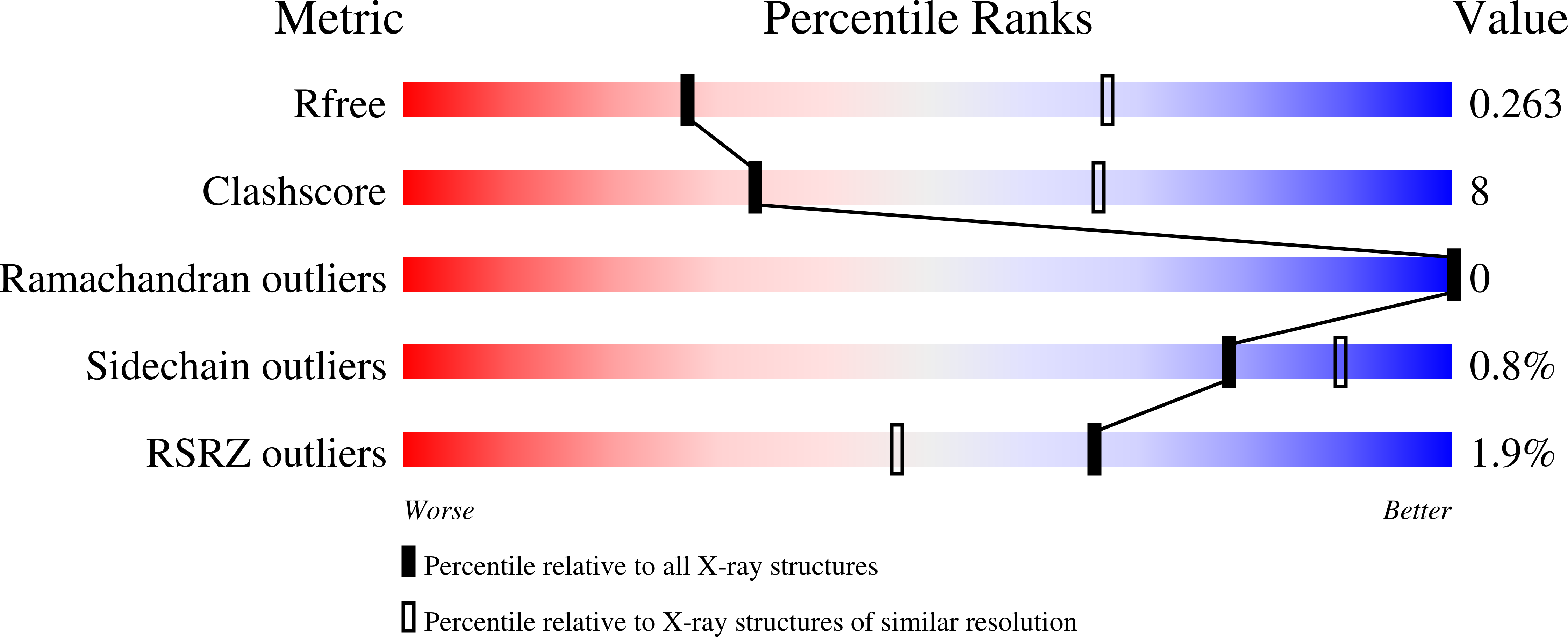
Resolution:
3.09 Å
R-Value Free:
0.26
R-Value Work:
0.23
R-Value Observed:
0.23
Space Group:
C 1 2 1
Macromolecular Entities
Structures with similar UniProt ID
Protein Blast
Polymer Type:polypeptide(L)
Molecule:Envelope glycoprotein E
Chain IDs:C (auth: A)
Chain Length:203
Number of Molecules:1
Biological Source:Human alphaherpesvirus 3
Polymer Type:polypeptide(L)
Molecule:Fab 1E12 Heavy Chain
Chain IDs:D (auth: B)
Chain Length:248
Number of Molecules:1
Biological Source:Homo sapiens
Polymer Type:polypeptide(L)
Molecule:Fab 1E12 Light Chain
Chain IDs:E (auth: C)
Chain Length:219
Number of Molecules:1
Biological Source:Homo sapiens
Polymer Type:polypeptide(L)
Molecule:Fab 1A2 Heavy Chain
Chain IDs:A (auth: H)
Chain Length:243
Number of Molecules:1
Biological Source:Homo sapiens
Polymer Type:polypeptide(L)
Molecule:Fab 1A2 Light Chain
Chain IDs:B (auth: L)
Chain Length:214
Number of Molecules:1
Biological Source:Homo sapiens
Ligand Molecules
Primary Citation
Structures of the Varicella Zoster Virus Glycoprotein E and Epitope Mapping of Vaccine-Elicited Antibodies.
Vaccines (Basel) 12 ? ? (2024)
PMID: 39460278 DOI: 10.3390/vaccines12101111

Abstact

Background: Varicella zoster virus (VZV) is the causative agent for chickenpox and herpes zoster (HZ, shingles). HZ is a debilitating disease affecting elderly and immunocompromised populations. Glycoprotein E (gE) is indispensable for viral replication and cell-to-cell spread and is the primary target for anti-VZV antibodies. Importantly, gE is the sole antigen in Shingrix, a highly efficacious, AS01B-adjuvanted vaccine approved in multiple countries for the prevention of HZ, yet the three-dimensional (3D) structure of gE remains elusive. Objectives: We sought to determine the structure of VZV gE and to understand in detail its interactions with neutralizing antibodies. Methods: We used X-ray crystallography and cryo-electron microscopy to elucidate structures of gE bound by recombinant Fabs of antibodies previously elicited through vaccination with Zostavax, a live, attenuated vaccine. Results: The 3D structures resolve distinct central and C-terminal antigenic domains, presenting an array of diverse conformational epitopes. The central domain has two beta-sheets and two alpha helices, including an IgG-like fold. The C-terminal domain exhibits 3 beta-sheets and an Ig-like fold and high structural similarity to HSV1 gE. Conclusions: gE from VZV-infected cells elicits a human antibody response with a preference for the gI binding domain of gE. These results yield insights to VZV gE structure and immunogenicity, provide a framework for future studies, and may guide the design of additional herpesvirus vaccine antigens. Teaser: Structures of varicella zoster virus glycoprotein E reveal distinct antigenic domains and define epitopes for vaccine-elicited human antibodies.

Legend

Protein

Chemical

Disease

Primary Citation of related structures