8V4H image
Entry Detail
PDB ID:
8V4H
Keywords:
Title:
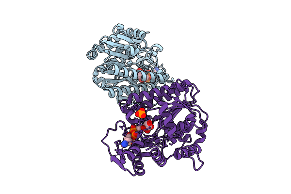
X-ray structure of the NADP-dependent reductase from Campylobacter jejuni responsible for the synthesis of CDP-glucitol in the presence of CDP-glucitol
Biological Source:
Source Organism:
PDB Version:
Deposition Date:
2023-11-29
Release Date:
2023-12-20
Method Details:
Experimental Method:
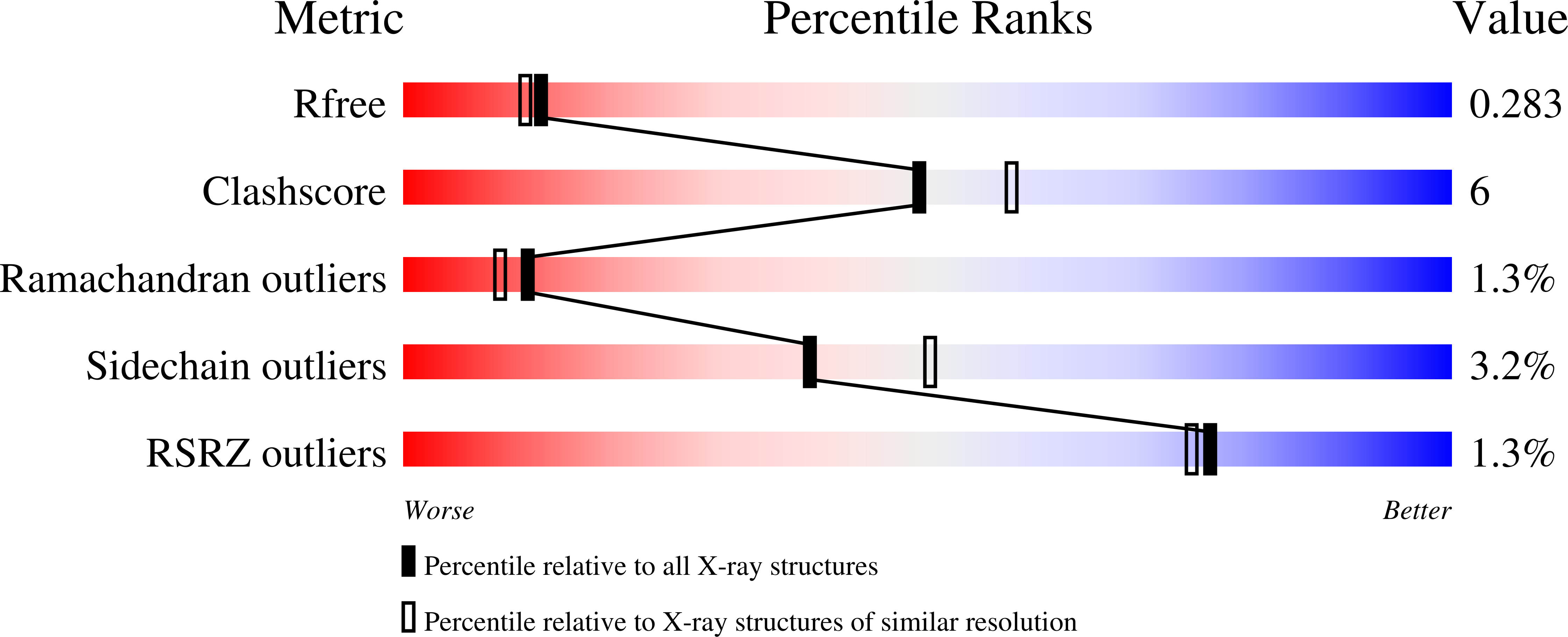
Resolution:
2.20 Å
R-Value Free:
0.28
R-Value Work:
0.20
R-Value Observed:
0.21
Space Group:
P 21 21 21
Macromolecular Entities
Polymer Type:polypeptide(L)
Description:Putative nucleotide sugar dehydratase
Chain IDs:A, B
Chain Length:356
Number of Molecules:2
Biological Source:Campylobacter jejuni
Primary Citation
Biosynthesis of Cytidine Diphosphate-6-d-Glucitol for the Capsular Polysaccharides of Campylobacter jejuni.
Biochemistry 63 699 710 (2024)
PMID: 38386885 DOI: 10.1021/acs.biochem.3c00706

Abstact

Campylobacter jejuni is a Gram-negative pathogenic bacterium commonly found in chickens and is the leading cause of human diarrheal disease worldwide. The various serotypes of C. jejuni produce structurally distinct capsular polysaccharides (CPSs) on the exterior surfaces of the cell wall. The capsular polysaccharide from C. jejuni serotype HS:5 is composed of a repeating sequence of d-glycero-d-manno-heptose and d-glucitol-6-phosphate. We previously defined the pathway for the production of d-glycero-d-manno-heptose in C. jejuni. Here, we elucidate the biosynthetic pathway for the assembly of cytidine diphosphate (CDP)-6-d-glucitol by the combined action of two previously uncharacterized enzymes. The first enzyme catalyzes the formation of CDP-6-d-fructose from cytidine triphosphate (CTP) and d-fructose-6-phosphate. The second enzyme reduces CDP-6-d-fructose with NADPH to generate CDP-6-d-glucitol. Using sequence similarity network (SSN) and genome neighborhood network (GNN) analyses, we predict that these pairs of proteins are responsible for the biosynthesis of CDP-6-d-glucitol and/or CDP-d-mannitol in the lipopolysaccharides (LPSs) and capsular polysaccharides in more than 200 other organisms. In addition, high resolution X-ray structures of the second enzyme are reported, which provide novel insight into the manner in which an open-chain nucleotide-linked sugar is harbored in an active site cleft.

Legend

Protein

Chemical

Disease

Primary Citation of related structures