8UZT image
Deposition Date 2023-11-16
Release Date 2024-08-21
Last Version Date 2024-10-02
Entry Detail
PDB ID:
8UZT
Title:
Mitochondrial single-stranded binding protein bound to DNA
Biological Source:
Source Organism:
Method Details:
Experimental Method:
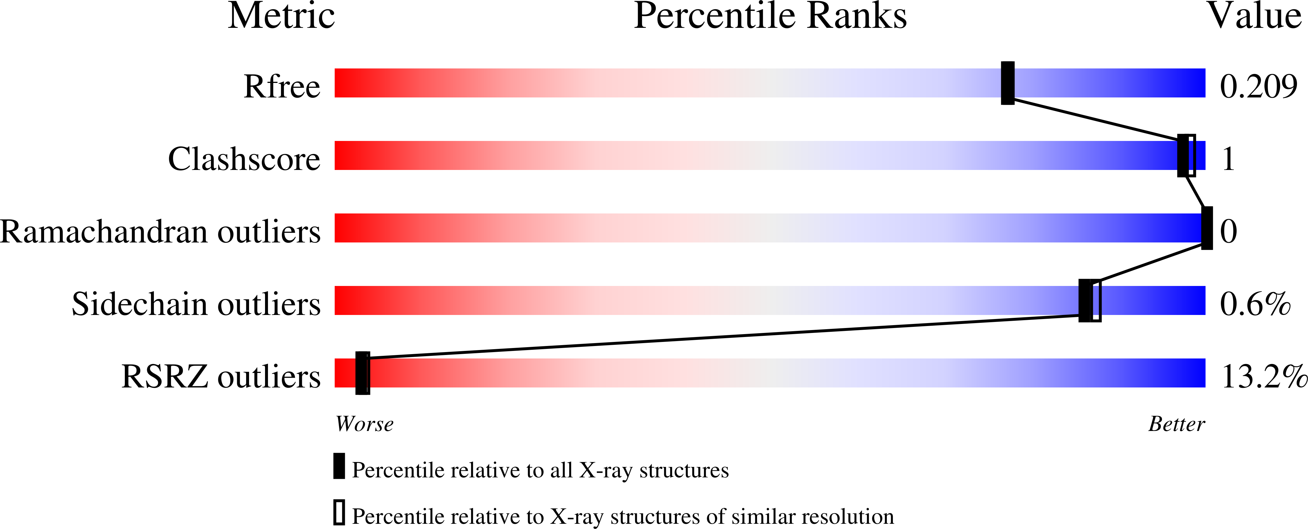
Resolution:
1.90 Å
R-Value Free:
0.20
R-Value Work:
0.18
R-Value Observed:
0.18
Space Group:
P 43 2 2
Macromolecular Entities
Polymer Type:polypeptide(L)
Molecule:Single-stranded DNA-binding protein, mitochondrial
Gene (Uniprot):SSBP1
Chain IDs:A (auth: B), B (auth: A), D (auth: C), E (auth: D)
Chain Length:154
Number of Molecules:4
Biological Source:Homo sapiens
Polymer Type:polydeoxyribonucleotide
Molecule:dTDNA
Chain IDs:C (auth: G)
Chain Length:20
Number of Molecules:1
Biological Source:synthetic construct
Primary Citation
Structures of the mitochondrial single-stranded DNA binding protein with DNA and DNA polymerase gamma.
Nucleic Acids Res. 52 10329 10340 (2024)
PMID: 39106165 DOI: 10.1093/nar/gkae670

Abstact

The mitochondrial single-stranded DNA (ssDNA) binding protein, mtSSB or SSBP1, binds to ssDNA to prevent secondary structures of DNA that could impede downstream replication or repair processes. Clinical mutations in the SSBP1 gene have been linked to a range of mitochondrial disorders affecting nearly all organs and systems. Yet, the molecular determinants governing the interaction between mtSSB and ssDNA have remained elusive. Similarly, the structural interaction between mtSSB and other replisome components, such as the mitochondrial DNA polymerase, Polγ, has been minimally explored. Here, we determined a 1.9-Å X-ray crystallography structure of the human mtSSB bound to ssDNA. This structure uncovered two distinct DNA binding sites, a low-affinity site and a high-affinity site, confirmed through site-directed mutagenesis. The high-affinity binding site encompasses a clinically relevant residue, R38, and a highly conserved DNA base stacking residue, W84. Employing cryo-electron microscopy, we confirmed the tetrameric assembly in solution and capture its interaction with Polγ. Finally, we derived a model depicting modes of ssDNA wrapping around mtSSB and a region within Polγ that mtSSB binds.

Legend

Protein

Chemical

Disease

Primary Citation of related structures