Maintenance Mode: Some services may turn out to be unavailable. We apologize for the inconvenience!
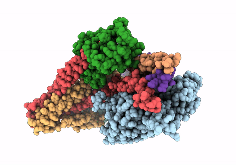
Cardiac muscle contraction occurs due to repetitive interactions between myosin thick and actin thin filaments (TF) regulated by Ca2+ levels, active cross-bridges, and cardiac myosin-binding protein C (cMyBP-C). The cardiac TF (cTF) has two nonequivalent strands, each comprised of actin, tropomyosin (Tm), and troponin (Tn). Tn shifts Tm away from myosin-binding sites on actin at elevated Ca2+ levels to allow formation of force-producing actomyosin cross-bridges. The Tn complex is comprised of three distinct polypeptides - Ca2+-binding TnC, inhibitory TnI, and Tm-binding TnT. The molecular mechanism of their collective action is unresolved due to lack of comprehensive structural information on Tn region of cTF. C1 domain of cMyBP-C activates cTF in the absence of Ca2+ to the same extent as rigor myosin. Here we used cryo-EM of native cTFs to show that cTF Tn core adopts multiple structural conformations at high and low Ca2+ levels and that the two strands are structurally distinct. At high Ca2+ levels, cTF is not entirely activated by Ca2+ but exists in either partially or fully activated state. Complete dissociation of TnI C-terminus is required for full activation. In presence of cMyBP-C C1 domain, Tn core adopts a fully activated conformation, even in absence of Ca2+. Our data provide a structural description for the requirement of myosin to fully activate cTFs and explain increased affinity of TnC to Ca2+ in presence of active cross-bridges. We suggest that allosteric coupling between Tn subunits and Tm is required to control actomyosin interactions.
Legend
Protein
Chemical
Disease