8UJA image
Deposition Date 2023-10-11
Release Date 2024-03-06
Last Version Date 2024-06-19
Entry Detail
PDB ID:
8UJA
Keywords:
Title:
T33-fn10 - Designed Tetrahedral Protein Cage Using Fragment-based Hydrogen Bond Networks
Biological Source:
Method Details:
Experimental Method:
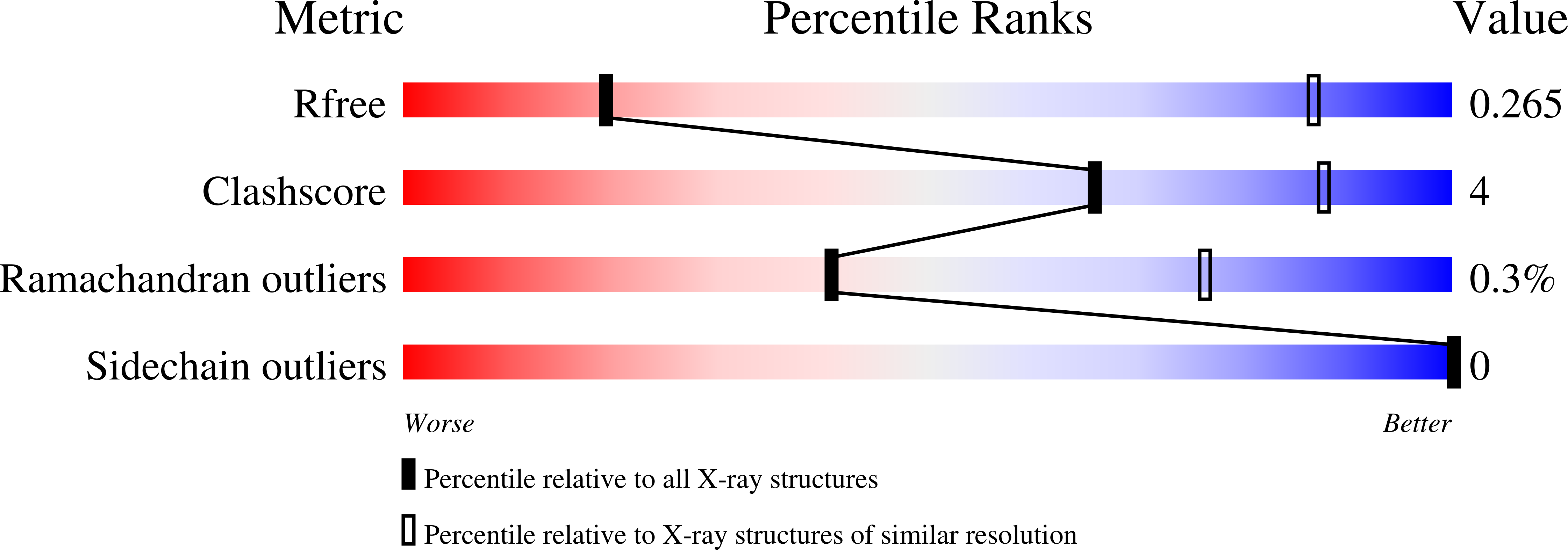
Resolution:
6.00 Å
R-Value Free:
0.24
R-Value Work:
0.21
R-Value Observed:
0.21
Space Group:
P 21 3
Macromolecular Entities
Polymer Type:polypeptide(L)
Molecule:T33-fn10: engineered DrsE like sulfur reductase
Chain IDs:A, C, E, G, I, K, M, O
Chain Length:108
Number of Molecules:8
Biological Source:Sulfurisphaera tokodaii str. 7
Structures with similar UniProt ID
Protein Blast
Polymer Type:polypeptide(L)
Molecule:T33-fn10: engineered enoyl-CoA hydratase/isomerase
Gene (Uniprot):Saro_3457
Chain IDs:B, D, F, H, J, L, N, P
Chain Length:258
Number of Molecules:8
Biological Source:Novosphingobium aromaticivorans DSM 12444
Ligand Molecules
Primary Citation
A suite of designed protein cages using machine learning and protein fragment-based protocols.
Structure 32 751 765.e11 (2024)
PMID: 38513658 DOI: 10.1016/j.str.2024.02.017

Abstact

Designed protein cages and related materials provide unique opportunities for applications in biotechnology and medicine, but their creation remains challenging. Here, we apply computational approaches to design a suite of tetrahedrally symmetric, self-assembling protein cages. For the generation of docked conformations, we emphasize a protein fragment-based approach, while for sequence design of the de novo interface, a comparison of knowledge-based and machine learning protocols highlights the power and increased experimental success achieved using ProteinMPNN. An analysis of design outcomes provides insights for improving interface design protocols, including prioritizing fragment-based motifs, balancing interface hydrophobicity and polarity, and identifying preferred polar contact patterns. In all, we report five structures for seven protein cages, along with two structures of intermediate assemblies, with the highest resolution reaching 2.0 Å using cryo-EM. This set of designed cages adds substantially to the body of available protein nanoparticles, and to methodologies for their creation.

Legend

Protein

Chemical

Disease

Primary Citation of related structures