Deposition Date
2023-10-05
Release Date
2024-06-19
Last Version Date
2024-11-06
Entry Detail
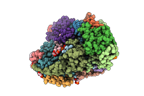
PDB ID:
8UGL
Keywords:
Title:
High resolution in-situ structure of complex IV in respiratory supercomplex
Biological Source:
Source Organism(s):
Sus scrofa (Taxon ID: 9823)
Method Details:
Experimental Method:
Resolution:
3.00 Å
Aggregation State:
PARTICLE
Reconstruction Method:
SINGLE PARTICLE