Deposition Date
2023-10-04
Release Date
2023-10-18
Last Version Date
2025-09-24
Entry Detail
PDB ID:
8UFL
Keywords:
Title:
Crystal Structure of SARS-Unique Domain (SUD) of Nsp3 from SARS coronavirus
Biological Source:
Source Organism(s):
Severe acute respiratory syndrome coronavirus (Taxon ID: 2901879)
Expression System(s):
Method Details:
Experimental Method:
Resolution:
2.51 Å
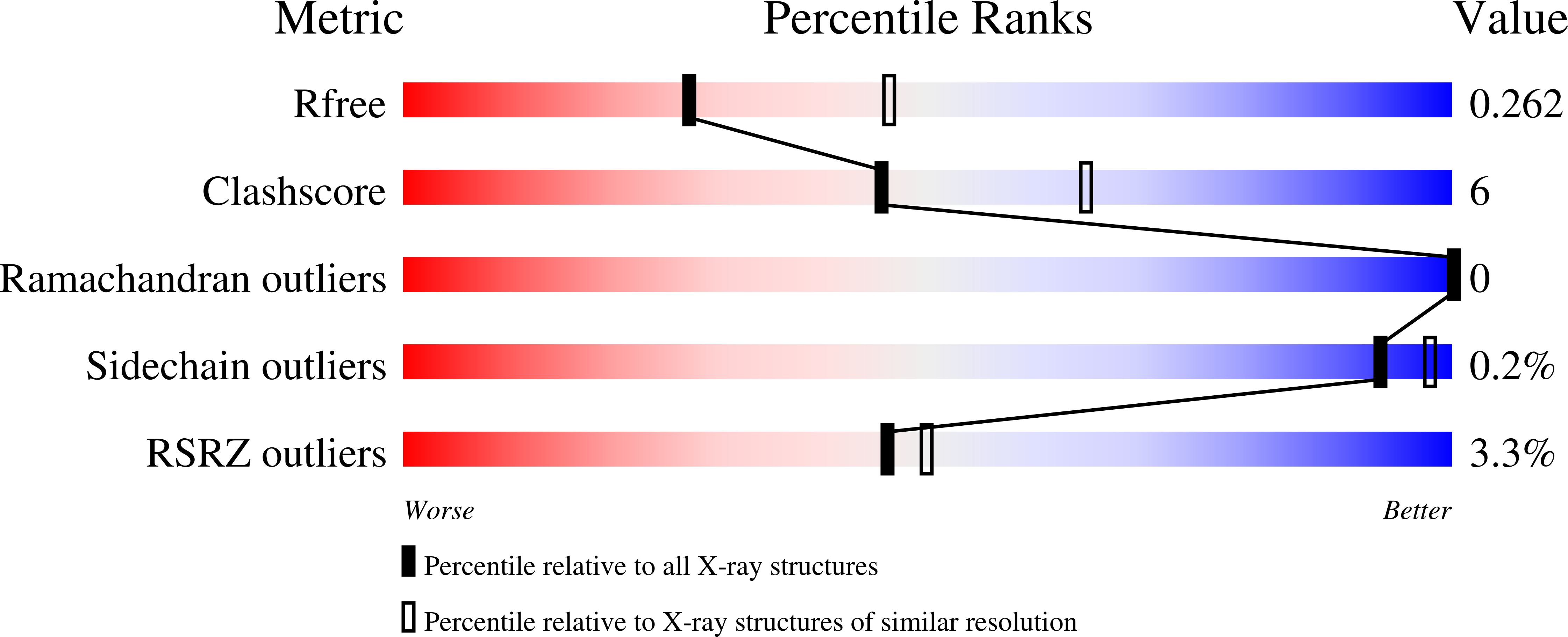
R-Value Free:
0.26
R-Value Work:
0.21
R-Value Observed:
0.21
Space Group:
P 21 21 21