8UEL image
Deposition Date 2023-10-01
Release Date 2023-12-13
Last Version Date 2024-11-20
Entry Detail
PDB ID:
8UEL
Keywords:
Title:
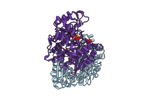
Crystal structure of enolase from Litopenaeus vannamei
Biological Source:
Source Organism:
Method Details:
Experimental Method:
Resolution:
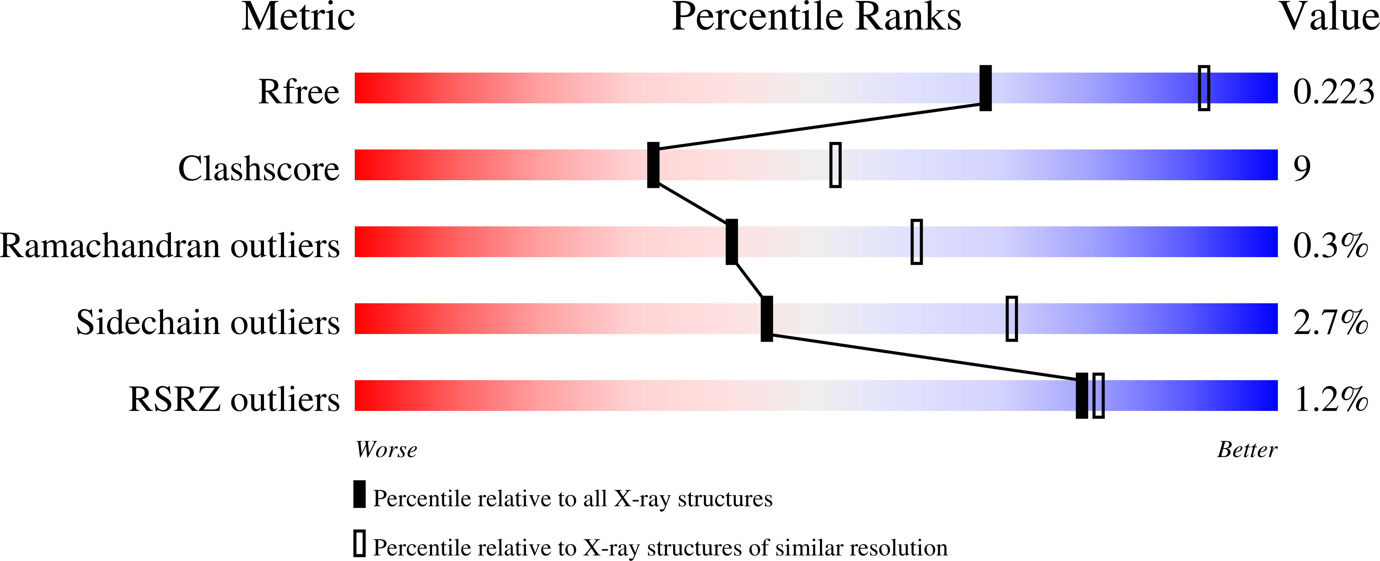
2.49 Å
R-Value Free:
0.22
R-Value Work:
0.16
R-Value Observed:
0.17
Space Group:
P 21 21 21
Macromolecular Entities
Structures with similar UniProt ID
Protein Blast
Polymer Type:polypeptide(L)
Molecule:Enolase
Chain IDs:A, B
Chain Length:433
Number of Molecules:2
Biological Source:Penaeus vannamei
Primary Citation
Characterization and Structural Analyses of Enolase from Shrimp ( Litopenaeus vannamei ).
J.Agric.Food Chem. 71 19783 19790 (2023)
PMID: 38033172 DOI: 10.1021/acs.jafc.3c07135

Abstact

Transcriptome analysis had recognized enolase from shrimp Litopenaeus vannamei (L. vannamei), which is termed LvEnolase, as one of the allergens, but its amino acid sequence and protein structure have been lacking. In this study, natural LvEnolase was isolated from L. vannamei and characterized for the first time. The full-length cDNA sequence of LvEnolase was effectively cloned, which encoded 434 amino acid residues. The crystal structure of LvEnolase was successfully determined at a resolution of 2.5 Å by X-ray crystallography (PDB: 8UEL). Notably, it was observed that near the active center, a loop exists in either an open or closed state, and the open loop was associated with the product release phase. Furthermore, enzyme activity assays were conducted to validate the catalytic capabilities of purified LvEnolase. These findings significantly enhance our comprehension of the enolase family and provide valuable support for delving into the functions and characteristics of LvEnolase.

Legend

Protein

Chemical

Disease

Primary Citation of related structures