8UEE image
Deposition Date 2023-10-01
Release Date 2023-12-27
Last Version Date 2024-07-10
Entry Detail
PDB ID:
8UEE
Keywords:
Title:
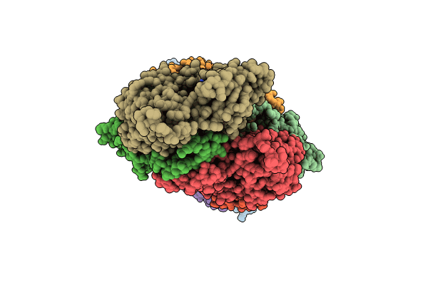
Atomic structure of Salmonella SipA/F-actin complex by cryo-EM
Biological Source:
Method Details:
Experimental Method:
Resolution:
3.20 Å
Aggregation State:
FILAMENT
Reconstruction Method:
HELICAL
Macromolecular Entities
Polymer Type:polypeptide(L)
Molecule:Cell invasion protein SipA
Gene (Uniprot):sipA
Chain IDs:H (auth: B), I (auth: C), J (auth: D), K (auth: A)
Chain Length:261
Number of Molecules:4
Biological Source:Salmonella enterica subsp. enterica serovar Typhimurium str. LT2
Polymer Type:polypeptide(L)
Molecule:Actin, alpha skeletal muscle
Gene (Uniprot):ACTA1
Chain IDs:A (auth: F), B (auth: H), C (auth: I), D (auth: J), E (auth: K), F (auth: L), G (auth: M)
Chain Length:375
Number of Molecules:7
Biological Source:Oryctolagus cuniculus
Modified Residue
Compound ID Chain ID Parent Comp ID Details 2D Image
HIC A HIS modified residue
Primary Citation
Stabilization of F-actin by Salmonella effector SipA resembles the structural effects of inorganic phosphate and phalloidin.
Structure 32 725 738.e8 (2024)
PMID: 38518780 DOI: 10.1016/j.str.2024.02.022

Abstact

Entry of Salmonella into host enterocytes relies on its pathogenicity island 1 effector SipA. We found that SipA binds to F-actin in a 1:2 stoichiometry with sub-nanomolar affinity. A cryo-EM reconstruction revealed that SipA's globular core binds at the groove between actin strands, whereas the extended C-terminal arm penetrates deeply into the inter-strand space, stabilizing F-actin from within. The unusually strong binding of SipA is achieved by a combination of fast association via the core and very slow dissociation dictated by the arm. Similar to Pi, BeF3, and phalloidin, SipA potently inhibited actin depolymerization by actin depolymerizing factor (ADF)/cofilin, which correlated with increased filament stiffness, supporting the hypothesis that F-actin's mechanical properties contribute to the recognition of its nucleotide state by protein partners. The remarkably strong binding to F-actin maximizes the toxin's effects at the injection site while minimizing global influence on the cytoskeleton and preventing pathogen detection by the host cell.

Legend

Protein

Chemical

Disease

Primary Citation of related structures